
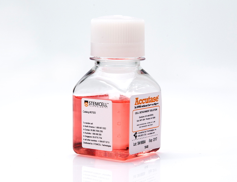
ACCUTASE™
Cell detachment solution

概要
A cell detachment solution of proteolytic and collagenolytic enzymes. Useful for the routine detachment of cells from standard tissue culture plasticware and adhesion coated plasticware. ACCUTASE™ does not contain mammalian or bacterial-derived products. Each lot of ACCUTASE™ is tested for sterility (by USP membrane filtration method), enzymatic activity (tested with synthetic chromagenic tetrapeptides) and cell detachment from tissue culture plastic.
Contains
• 1X ACCUTASE™ enzymes in Dulbecco’s phosphate-buffered saline (PBS)
• 0.5 mM EDTA•4Na
• 3 mg/L Phenol red
• 0.5 mM EDTA•4Na
• 3 mg/L Phenol red
Subtype
Enzymatic
Cell Type
Neural Cells, PSC-Derived, Pluripotent Stem Cells
Species
Human, Mouse, Rat, Non-Human Primate, Other
Area of Interest
Neuroscience, Stem Cell Biology
技术资料
| Document Type | 产品名称 | Catalog # | Lot # | 语言 |
|---|---|---|---|---|
| Product Information Sheet | ACCUTASE™ | 07922, 07920 | All | English |
| Safety Data Sheet | ACCUTASE™ | 07920 | All | English |
数据及文献
Publications (53)
Biomaterials 2018 JUN
Substrate stiffness modulates the multipotency of human neural crest derived ectomesenchymal stem cells via CD44 mediated PDGFR signaling.
Abstract
Abstract
Mesenchymal stem cells (MSCs) have been isolated from various mesodermal and ectodermal tissues. While the phenotypic and functional heterogeneity of MSCs stemming from their developmental origins has been acknowledged, the genetic and environmental factors underpinning these differences are not well-understood. Here, we investigated whether substrate stiffness mediated mechanical cues can directly modulate the development of ectodermal MSCs (eMSCs) from a precursor human neural crest stem cell (NCSC) population. We showed that NCSC-derived eMSCs were transcriptionally and functionally distinct from mesodermal bone marrow MSCs. eMSCs derived on lower substrate stiffness specifically increased their expression of the MSC marker, CD44 in a Rho-ROCK signaling dependent manner, which resulted in a concomitant increase in the eMSCs' adipogenic and chondrogenic differentiation potential. This mechanically-induced effect can only be maintained for short-term upon switching back to a stiff substrate but can be sustained for longer-term when the eMSCs were exclusively maintained on soft substrates. We also discovered that CD44 expression modulated eMSC self-renewal and multipotency via the downregulation of downstream platelet-derived growth factor receptor beta (PDGFRbeta$) signaling. This is the first instance demonstrating that substrate stiffness not only influences the differentiation trajectories of MSCs but also their derivation from upstream progenitors, such as NCSCs.
Current protocols in cell biology 2018 JUN
Transcription Factor-Mediated Differentiation of Human iPSCs into Neurons.
Abstract
Abstract
Accurate modeling of human neuronal cell biology has been a long-standing challenge. However, methods to differentiate human induced pluripotent stem cells (iPSCs) to neurons have recently provided experimentally tractable cell models. Numerous methods that use small molecules to direct iPSCs into neuronal lineages have arisen in recent years. Unfortunately, these methods entail numerous challenges, including poor efficiency, variable cell type heterogeneity, and lengthy, expensive differentiation procedures. We recently developed a new method to generate stable transgenic lines of human iPSCs with doxycycline-inducible transcription factors at safe-harbor loci. Using a simple two-step protocol, these lines can be inducibly differentiated into either cortical (i3 Neurons) or lower motor neurons (i3 LMN) in a rapid, efficient, and scalable manner (Wang et al., 2017). In this manuscript, we describe a set of protocols to assist investigators in the culture and genetic engineering of iPSC lines to enable transcription factor-mediated differentiation of iPSCs into i3 Neurons or i3 LMNs, and we present neuronal culture conditions for various experimental applications. {\textcopyright} 2018 by John Wiley & Sons, Inc.
Molecular neurodegeneration 2018
iPSC-derived familial Alzheimer's PSEN2 N141I cholinergic neurons exhibit mutation-dependent molecular pathology corrected by insulin signaling.
Abstract
Abstract
BACKGROUND Type 2 diabetes (T2D) is a recognized risk factor for the development of cognitive impairment (CI) and/or dementia, although the exact nature of the molecular pathology of T2D-associated CI remains obscure. One link between T2D and CI might involve decreased insulin signaling in brain and/or neurons in either animal or postmortem human brains as has been reported as a feature of Alzheimer's disease (AD). Here we asked if neuronal insulin resistance is a cell autonomous phenomenon in a familial form of AD. METHODS We have applied a newly developed protocol for deriving human basal forebrain cholinergic neurons (BFCN) from skin fibroblasts via induced pluripotent stem cell (iPSC) technology. We generated wildtype and familial AD mutant PSEN2 N141I (presenilin 2) BFCNs and assessed if insulin signaling, insulin regulation of the major AD proteins Abeta$ and/or tau, and/or calcium fluxes is altered by the PSEN2 N141I mutation. RESULTS We report herein that wildtype, PSEN2 N141I and CRISPR/Cas9-corrected iPSC-derived BFCNs (and their precursors) show indistinguishable insulin signaling profiles as determined by the phosphorylation of canonical insulin signaling pathway molecules. Chronic insulin treatment of BFCNs of all genotypes led to a reduction in the Abeta$42/40 ratio. Unexpectedly, we found a CRISPR/Cas9-correctable effect of PSEN2 N141I on calcium flux, which could be prevented by chronic exposure of BFCNs to insulin. CONCLUSIONS Our studies indicate that the familial AD mutation PSEN2 N141I does not induce neuronal insulin resistance in a cell autonomous fashion. The ability of insulin to correct calcium fluxes and to lower Abeta$42/40 ratio suggests that insulin acts to oppose an AD-pathophysiology. Hence, our results are consistent with a potential physiological role for insulin as a mediator of resilience by counteracting specific metabolic and molecular features of AD.
Frontiers in Molecular Neuroscience 2018
Patch-Seq Protocol to Analyze the Electrophysiology, Morphology and Transcriptome of Whole Single Neurons Derived From Human Pluripotent Stem Cells
Abstract
Abstract
The human brain is composed of a complex assembly of about 171 billion heterogeneous cellular units (86 billion neurons and 85 billion non-neuronal glia cells). A comprehensive description of brain cells is necessary to understand the nervous system in health and disease. Recently, advances in genomics have permitted the accurate analysis of the full transcriptome of single cells (scRNA-seq). We have built upon such technical progress to combine scRNA-seq with patch-clamping electrophysiological recording and morphological analysis of single human neurons in vitro. This new powerful method, referred to as Patch-seq, enables a thorough, multimodal profiling of neurons and permits us to expose the links between functional properties, morphology, and gene expression. Here, we present a detailed Patch-seq protocol for isolating single neurons from in vitro neuronal cultures. We have validated the Patch-seq whole-transcriptome profiling method with human neurons generated from embryonic and induced pluripotent stem cells (ESCs/iPSCs) derived from healthy subjects, but the procedure may be applied to any kind of cell type in vitro. Patch-seq may be used on neurons in vitro to profile cell types and states in depth to unravel the human molecular basis of neuronal diversity and investigate the cellular mechanisms underlying brain disorders.
Cell stem cell 2017 APR
Human iPSC-Derived Cerebral Organoids Model Cellular Features of Lissencephaly and Reveal Prolonged Mitosis of Outer Radial Glia.
Abstract
Abstract
Classical lissencephaly is a genetic neurological disorder associated with mental retardation and intractable epilepsy, and Miller-Dieker syndrome (MDS) is the most severe form of the disease. In this study, to investigate the effects of MDS on human progenitor subtypes that control neuronal output and influence brain topology, we analyzed cerebral organoids derived from control and MDS-induced pluripotent stem cells (iPSCs) using time-lapse imaging, immunostaining, and single-cell RNA sequencing. We saw a cell migration defect that was rescued when we corrected the MDS causative chromosomal deletion and severe apoptosis of the founder neuroepithelial stem cells, accompanied by increased horizontal cell divisions. We also identified a mitotic defect in outer radial glia, a progenitor subtype that is largely absent from lissencephalic rodents but critical for human neocortical expansion. Our study, therefore, deepens our understanding of MDS cellular pathogenesis and highlights the broad utility of cerebral organoids for modeling human neurodevelopmental disorders.
Annals of hematology 2016 OCT
Generation of induced pluripotent stem cells as a potential source of hematopoietic stem cells for transplant in PNH patients.
Abstract
Abstract
Paroxysmal nocturnal hemoglobinuria (PNH) is an acquired hemolytic anemia caused by lack of CD55 and CD59 on blood cell membrane leading to increased sensitivity of blood cells to complement. Hematopoietic stem cell transplantation (HSCT) is the only curative therapy for PNH, however, lack of HLA-matched donors and post-transplant complications are major concerns. Induced pluripotent stem cells (iPSCs) derived from patients are an attractive source for generating autologous HSCs to avoid adverse effects resulting from allogeneic HSCT. The disease involves only HSCs and their progeny; therefore, other tissues are not affected by the mutation and may be used to produce disease-free autologous HSCs. This study aimed to derive PNH patient-specific iPSCs from human dermal fibroblasts (HDFs), characterize and differentiate to hematopoietic cells using a feeder-free protocol. Analysis of CD55 and CD59 expression was performed before and after reprogramming, and hematopoietic differentiation. Patients' dermal fibroblasts expressed CD55 and CD59 at normal levels and the normal expression remained after reprogramming. The iPSCs derived from PNH patients had typical pluripotent properties and differentiation capacities with normal karyotype. After hematopoietic differentiation, the differentiated cells expressed early hematopoietic markers (CD34 and CD43) with normal CD59 expression. The iPSCs derived from HDFs of PNH patients have normal levels of CD55 and CD59 expression and hold promise as a potential source of HSCs for autologous transplantation to cure PNH patients.