
Calcitriol
Vitamin D receptor activator


概要
Calcitriol is synthesized from 25-hydroxy Vitamin D3, the principal circulating form of Vitamin D, via hydroxylation in the kidney. The main physiologic effects of calcitriol are to increase the absorption of calcium at the level of the intestinal epithelium, and to increase the mineralization of bone via the direct stimulation of osteoblasts (Portale et al.)
DIFFERENTIATION
· Induces differentiation of human osteoblasts, alone or in combination with TGFβ (Ingram et al.; Kassem et al.; Wergedal et al.).
· Induces differentiation of chicken embryonic chondrocytes (Gerstenfeld et al.; Tsonis).
· Enhances differentiation of human keratinocytes when grown in the presence of high calcium concentrations (Itin et al.).
Alternative Names
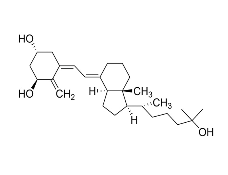
1α,25-dihydroxy vitamin D₃
Cell Type
Chondrocytes, Keratinocytes, Mesenchymal Stem and Progenitor Cells, Osteoblasts
Species
Human, Mouse, Rat, Non-Human Primate, Other
Application
Differentiation
Area of Interest
Stem Cell Biology
CAS Number
32222-06-3
Chemical Formula
C₂₇H₄₄O₃
Molecular Weight
416.6 g/mol
Purity
≥ 97%
Pathway
Vitamin D
Target
Vitamin D Receptor
技术资料
| Document Type | 产品名称 | Catalog # | Lot # | 语言 |
|---|---|---|---|---|
| Product Information Sheet | Calcitriol | 72412 | All | English |
| Safety Data Sheet | Calcitriol | 72412 | All | English |
数据及文献
Publications (8)
European journal of clinical investigation 2000 MAY
Production and action of transforming growth factor-beta in human osteoblast cultures: dependence on cell differentiation and modulation by calcitriol.
Abstract
Abstract
BACKGROUND: Transforming growth factor beta (TGF-beta) plays an important role in skeletal remodelling. However, few studies have examined its effects on cultured human osteoblasts. Our aim is to characterise the biological effects of TGF-beta1 on human osteoblasts and to examine the interaction between TGF-beta1 and calcitriol. DESIGN: In vitro study employing two models of normal human osteoblasts: human bone marrow stromal cells [hMS/(OB)] containing osteoprogenitor cells and trabecular bone osteoblasts (hOB), which are mature osteoblasts. A reverse-transcriptase-polymerase-chain-reaction assay was employed to measure steady state mRNA levels of TGF-beta(s) isoforms and receptors. Effects of short-term treatment of TGF-beta1 on osteoblast proliferation and differentiation markers were assessed. The effect of cotreatment of calcitriol (10-8 M) and TGF-beta1 on osteoblast differentiation was also determined. RESULTS: Both hMS(OB) and hOB cells expressed mRNA transcripts of TGF-beta1, TGF-beta2, TGF-beta 3, TGF-beta type I and type II receptors. TGF-beta 1 stimulated osteoblast proliferation in hMS(OB) and in hOB cultures. In hOB cultures, TGF-beta1 stimulated AP production and cotreatment with calcitriol induced a synergistic increase in AP levels to 250 +/- 61% of calcitriol-treated controls. Effects of TGF-beta1 and calcitriol were less pronounced in hMS(OB) cultures. TGF-beta1 inhibited collagen type I production in hMS(OB) cells and these effects were abolished in presence of calcitriol. In presence of calcitriol, TGF-beta1 increased collagen type I production in hOB cells. In both hOB and hMS(OB) cultures, TGF-beta1 inhibited osteocalcin production. CONCLUSIONS: TGF-beta increases osteoblastic cell proliferation irrespective of the differentiation state. In presence of calcitriol, it initiates osteoblast cell differentiation and matrix formation. As TGF-beta inhibits osteocalcin production, other factors are necessary for inducing terminal differentiation of osteoblasts. The observed effects of TGF-beta on human osteoblasts in vitro may represent important regulatory steps in controlling osteoblast cell proliferation and differentiation in vivo.
Endocrinology 1994 NOV
Effects of vitamin D metabolites on proliferation and differentiation of cultured human epidermal keratinocytes grown in serum-free or defined culture medium.
Abstract
Abstract
We examined the effects of 1,25-dihydroxyvitamin D3 [1,25(OH)2D3], 25-hydroxyvitamin D3 (25OHD3), and vitamin D3 on human keratinocyte proliferation and differentiation in a serum-free or defined culture system. Concentrations greater than 10(-8) M 1,25-(OH)2D3 or 10(-7) M 25(OH)2D3 caused marked inhibition of cell growth. Growth inhibition with high doses of 1,25-(OH)2D3 was not stringent, but was mainly exerted in the G1 phase of the cell cycle. Early release from the cell cycle block restored the proliferation of human keratinocytes. The calcium concentration in the medium had no significant effect on the antiproliferative action of 1,25-(OH)2D3, 25OHD3, and vitamin D3. We also show that human keratinocyte proliferation is enhanced at doses of 1,25-(OH)2D3 and 25OH2D3 of 10(-9) M or less. Enhanced proliferation of human keratinocytes with physiological concentrations of 1,25-(OH)2D3 could only be shown in fully defined medium that contained no vitamin D3, related sterols, or bovine pituitary extract. Human keratinocyte differentiation was enhanced with higher doses of 1,25-(OH)2D3 when cells were grown in the presence of high calcium concentrations. These studies demonstrate that the lower, physiological concentrations of vitamin D3 metabolites are capable of stimulating the proliferation of epidermal keratinocytes grown under selected conditions that eliminate confounding or unidentified medium culture factors. Vitamin D3 metabolites are shown to exert mitogenic trophic effects in cultured human epithelial cells similar to their established activities in vivo.
Differentiation; research in biological diversity 1994 JAN
Effects of transforming growth factor beta (TGF beta) and 1,25 dihydroxyvitamin D3 on the function, cytochemistry and morphology of normal human osteoblast-like cells.
Abstract
Abstract
Individually, transforming growth factor beta (TGF beta) and 1,25-dihydroxyvitamin D3 (1,25(OH)2D3) alter the growth and differentiation of normal and transformed osteoblast-like (OB) cells. Although recent evidence suggests interactions between TGF beta and 1,25(OH)2D3 may occur, little is known of the individual or combined effects of these hormones on the expression of the osteoblast phenotype at the cytochemical and biochemical levels in normal human OB (hOB) cells. Primary cultures of hOBs were treated with TGF beta (0.001-10 ng/ml) and 1,25(OH)2D3 (0.1 pM-100 nM) either alone or in combination. TGF beta and 1,25(OH)2D3 stimulated spindle-shaped cells to become stellate in appearance and increased the number of cytoplasmic processes. TGF beta increased 3H-thymidine incorporation and 1,25(OH)2D3 reduced this effect. Conversely, procollagen type-I synthesis and secretion were increased in a dose-dependent manner in the presence of TGF beta but were not significantly affected in the presence of 1,25(OH)2D3. TGF beta and 1,25(OH)2D3 each marginally increased alkaline phosphatase (ALP) activity, but the combination synergistically increased ALP activity in a dose- and time-dependent manner at the cytochemical and biochemical level (three to tenfold over vehicle controls; n = 12). In contrast, TGF beta reduced 1,25(OH)2D3-stimulated osteocalcin secretion. These data suggest that TGF beta stimulates hOB cells to actively produce collagen matrix and proliferate. The combination of TGF beta and 1,25(OH)2D3, however, produces a synergistic increase in ALP activity and maintenance of collagen synthesis. 1,25(OH)2D3 stimulation may induce cells to advance to an endstage where cell proliferation is reduced and osteocalcin expression is promoted. Interactions between TGF beta and 1,25(OH)2D3 may represent important steps in the regulation of osteoblast differentiation and matrix production.
Metabolism: clinical and experimental 1992 JAN
Differentiation of normal human bone cells by transforming growth factor-beta and 1,25(OH)2 vitamin D3.
Abstract
Abstract
To investigate the role of transforming growth factor-beta 1 (TGF beta) in bone metabolism, the effects of this agent on the differentiation characteristics of human bone cells were studied in vitro. Human bone cells were isolated from femoral head samples by collagenase digestion. Differentiation characteristics included alkaline phosphatase activity, osteocalcin production, and mRNA levels for alkaline phosphatase, type I alpha 2-procollagen, and osteocalcin. The effect of TGF beta on alkaline phosphatase was not constant, but varied with the incubation conditions. At high cell density and in the presence of serum, TGF beta decreased alkaline phosphatase activity. However, at low cell density and under serum-free conditions, TGF beta stimulated alkaline phosphatase activity. The addition of 1,25(OH)2 vitamin D3 also stimulated alkaline phosphatase. The combination of the two agents gave a greater increase in activity than the sum of the activities when the two agents were given alone. The percentage of cells that stain positively for alkaline phosphatase changed in parallel with the change in specific activity. The percentage of positive cells increased from 17% to 64%, while the specific activity increased from 22 to 169 mU/mg protein. To investigate the mechanism of this stimulation, mRNA levels were measured at 24 hours. Individually, TGF beta and 1,25(OH)2D3 increased message levels for alkaline phosphatase and type I procollagen, but the greatest effect was produced by the combination of the two factors. 1,25(OH)2D3 increased osteocalcin mRNA levels, but TGF beta markedly inhibited this stimulation. TGF beta also inhibited production of osteocalcin by the human bone cells. TGF beta appears to modulate differentiation of human bone cells in combination with 1,25(OH)2D3 and other factors.
Developmental biology 1991 JAN
1,25-Dihydroxyvitamin D3 stimulates chondrogenesis of the chick limb bud mesenchymal cells.
Abstract
Abstract
Vitamin D has been known to be important for skeletal development and growth but the mechanism whereby it affects these processes is not well understood. We report now that the hormonal metabolite of vitamin D3, namely 1,25-dihydroxyvitamin D3, stimulates chondrogenesis in cultures of stage 24 chick embryo limb bud mesenchymal cells, as evidenced by morphologic changes as well as by increased transcription of collagen type II and core protein genes. This effect appears to be specific to 1,25(OH)2D3 since 24,25(OH)2D3 or D3 does not influence chondrogenesis in this system, and is probably mediated via the specific 1,25(OH)2D3 receptor protein which is undetectable in untreated cells but appears following exposure to the hormone.
Endocrinology 1990 MAR
Effect of 1,25-dihydroxyvitamin D3 on induction of chondrocyte maturation in culture: extracellular matrix gene expression and morphology.
Abstract
Abstract
Chondrocytes, derived from a tissue that remains as permanent hyaline cartilage in vivo (embryonic chicken caudal sterna) were treated with 10(-8) to 10(-8) M 1,25-dihydroxyvitamin D3 [1,25(OH)2D3]. These nonadherent rounded chondrocytes acquired an adherent, polygonal morphology in a dose-dependent fashion with 1,25(OH)2D3 treatment. During the first 4 days of 1,25(OH)2D3 treatment cell flattening was associated with a 10-fold increase in beta-actin and fibronectin and their corresponding messenger RNAs (mRNAs). After adherence over the 12 days of continuous hormone treatment, a 2- to 4-fold increase in DNA synthesis and DNA accumulation were observed for the highest hormone dose (10(-8) M). Over the same time course total collagen synthesis decreased 35-50% primarily due to decreased type II collagen synthesis, which accompanied comparable decreases in its mRNA. In contrast, both alpha 1(I) and alpha 2(I) showed a continuous 5- to 10-fold increase; however, type I collagen protein synthesis remained undetectable, indicating translational control of the type I collagen synthesis. alpha 1(X) mRNAs showed a 2- 3-fold increase after 12 days of hormone treatment, and its polypeptide was clearly detected by sodium dodecyl sulfate polyacrylamide gel analysis. Type IX collagen synthesis showed a 2-fold increase in synthesis and its mRNA levels during the first 4 days of 1,25(OH)2D3 treatment but thereafter had levels comparable to control cultures. Analysis of proteoglycan synthesis and core protein mRNA levels showed there was a 2-fold increase in core protein mRNAs while proteoglycan synthesis, as assessed by 35S incorporation, showed only a 10-20% increase. Direct hormone effects vs. those secondary to altered cellular morphology were determined by blocking cell adherence by growth of the 1,25(OH)2D3-treated cultures on bacteriological petri dishes. All of the observed effects on cytoskeletal and collagen mRNAs were blocked except the elevations observed in proteoglycan core protein and alpha 1(IX) mRNAs. DNA contents in hormone-treated cultures also remained elevated. These results suggest that 1,25(OH)2D3 both activates and suppresses specific genes, promoting chondrocyte maturation toward a more hypertrophic phenotype. However, prevention of the initial morphological alterations that are induced by 1,25(OH)2D3 blocks many of the subsequent changes in connective tissue expression.