
Paclitaxel
Inhibitor of microtubule formation


概要
Paclitaxel is a diterpene alkaloid originally isolated from the bark of the Pacific Yew tree (Taxus brevifolia). It binds to and stabilizes microtubules, preventing their reorganization during cell division, which leads to cell cycle arrest. Paclitaxel has antitumorigenic properties and has been used as a chemotherapeutic compound (Rowinsky et al.). Many pathways have been implicated in Paclitaxel-induced apoptosis, including c-Jun N-terminal kinase/stress-activated protein kinase (JNK/SAPK), p38 mitogen-activated protein kinase (MAPK), and protein kinase A (PKA; Wang et al.; Reshkin et al.).
DIFFERENTIATION
· Inhibits initiation and outgrowth of neurites in vitro, through microtubule polymerization (Letourneau & Ressler).
CANCER RESEARCH
· Inhibits tumor cell growth in a variety of cancer cell lines including cervical (HeLa), lung (A549), breast (MCF-7), colon (HT-29), ovarian (OVG-1), and pancreatic (PC-Sh) carcinomas (Liebmann et al.).
· Induces abnormal multipolar spindle formation, inducing cell cycle arrest at prophase and G1 in various human cell cancer lines (Woods et al.).
· Initiates apoptosis of cancer cells through multiple mechanisms involving: p53-dependent and -independent pathways, B-cell CLL/lymphoma 2 (BCL-2) family members, cyclin-dependent kinases, p38 MAPK, PKA, and JNK/SAPK (Wang et al.; Reshkin et al.).
· Induces cyclin inhibitor p21 in MCF7 and PC3M human cancer cell lines by a mechanism dependent on the activation of RAF-1 (Blagosklonny et al.).
Alternative Names
NSC 125973
Cell Type
Cancer Cells and Cell Lines, Leukemia/Lymphoma Cells, Neurons
Species
Human, Mouse, Rat, Non-Human Primate, Other
Area of Interest
Cancer Research, Neuroscience
CAS Number
33069-62-4
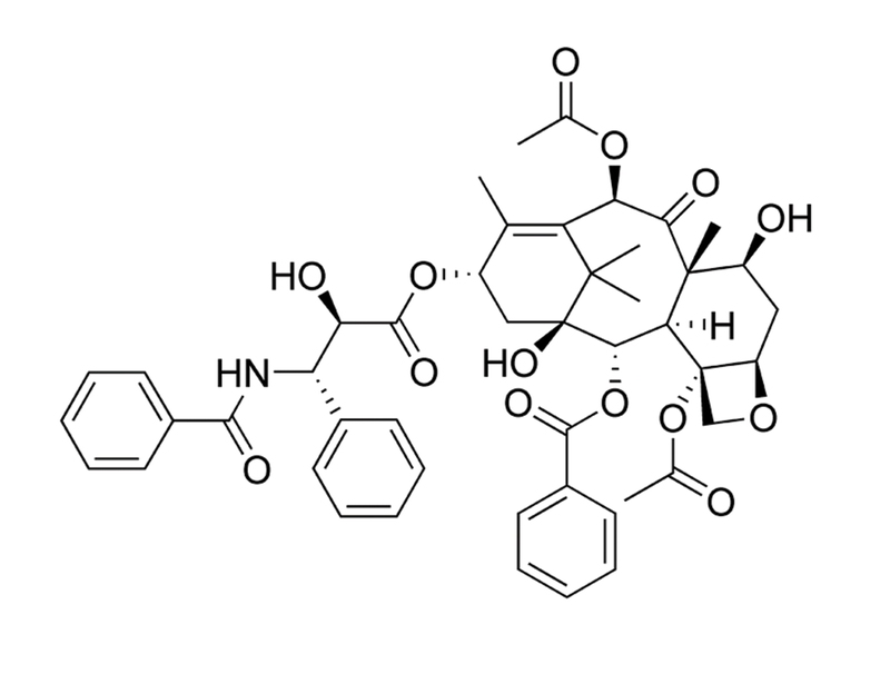
Chemical Formula
C₄₇H₅₁NO₁₄
Molecular Weight
853.9 g/mol
Purity
≥ 98%
技术资料
| Document Type | 产品名称 | Catalog # | Lot # | 语言 |
|---|---|---|---|---|
| Product Information Sheet | Paclitaxel | 73312, 73314 | All | English |
| Safety Data Sheet | Paclitaxel | 73312, 73314 | All | English |
数据及文献
Publications (7)
Clinical cancer research : an official journal of the American Association for Cancer Research 2003
Paclitaxel induces apoptosis via protein kinase A- and p38 mitogen-activated protein-dependent inhibition of the Na+/H+ exchanger (NHE) NHE isoform 1 in human breast cancer cells.
Abstract
Abstract
PURPOSE: The molecular signal components essential to paclitaxel-dependent apoptosis in breast cancers are potential targets for combined therapy. However, the signal mechanisms underlying paclitaxel action still need to be better defined. EXPERIMENTAL DESIGN: In a breast cancer cell line, pharmacological agents and transient transfection with dominant interfering and constitutive active mutants were used to identify the signal transduction module involved in the regulation of paclitaxel-induced apoptosis and to evaluate its potential as a therapeutic target. RESULTS: In MDA-MB-435 cells, paclitaxel treatment stimulated the activity of both protein kinase A and p38, and inhibited the activity of the Na(+)/H(+) exchanger isoform 1 (NHE1) with similar IC(50) concentrations as for its activation of apoptosis. Activation and inhibition experiments demonstrated that protein kinase A and p38 participate sequentially upstream of the NHE1 in regulating the paclitaxel-induced apoptotic pathway. Importantly, concurrent specific inhibition of the NHE1 with paclitaxel treatment resulted in a synergistic induction of apoptosis and a reduction in the paclitaxel IC(50) for apoptosis. This sensitization of paclitaxel apoptotic action by specific inhibition of NHE1 was verified in breast cancer cell lines with different paclitaxel sensitivity. CONCLUSIONS: We have, for the first time, identified NHE1 as an essential component of paclitaxel-induced apoptosis in breast cancer cells and, importantly, identified that simultaneous inhibition of the NHE1 results in a synergistic potentiation of low-dose paclitaxel apoptotic action. As specific NHE1 inhibitors have finished Phase II/Phase III clinical trials for myocardial protection, there is the possibility for a rapid biological translation of this novel therapeutic strategy to a clinical setting.
Cancer 2000
Paclitaxel-induced cell death: where the cell cycle and apoptosis come together.
Abstract
Abstract
BACKGROUND: Compelling evidence indicates that paclitaxel kills cancer cells through the induction of apoptosis. Paclitaxel binds microtubules and causes kinetic suppression (stabilization) of microtubule dynamics. The consequent arrest of the cell cycle at mitotic phase has been considered to be the cause of paclitaxel-induced cytotoxicity. However, the biochemical events, downstream from paclitaxel's binding to microtubules, that lead to apoptosis are not well understood. METHODS: The authors examined recent scientific literature about the mechanisms by which paclitaxel exerts cytotoxicity. RESULTS: In addition to an arrest of the cell cycle at the mitotic phase in paclitaxel-treated cells, recent discoveries of activation of signaling molecules by paclitaxel and paclitaxel-induced transcriptional activation of various genes indicate that paclitaxel initiates apoptosis through multiple mechanisms. The checkpoint of mitotic spindle assembly, aberrant activation of cyclin-dependent kinases, and the c-Jun N-terminal kinase/stress-activated protein kinase (JNK/SAPK) are shown to be involved in paclitaxel-induced apoptosis. Consistent with observations that microtubules of different status (e.g., cytoskeletal microtubules vs. mitotic spindles) have different sensitivity to paclitaxel, the concentration of paclitaxel appears to be the major determinant of its apoptogenic mechanisms. CONCLUSIONS: Advances in research of the cell cycle and apoptosis have extended our understanding of the mechanisms of paclitaxel-induced cell death. Further elucidation of resistance and enhancement of paclitaxel-induced apoptosis should expedite the development of better paclitaxel-based regimens for cancer therapy.
Molecular medicine (Cambridge, Mass.) 1995
Taxol-induced mitotic block triggers rapid onset of a p53-independent apoptotic pathway.
Abstract
Abstract
BACKGROUND: At therapeutic concentrations, the antineoplastic agent taxol selectively perturbs mitotic spindle microtubules. Taxol has recently been shown to induce apoptosis, similar to the mechanism of cell death induced by other antineoplastic agents. However, taxol has shown efficacy against drug-refractory cancers, raising the possibility that this pharmacological agent may trigger an alternative apoptotic pathway. MATERIALS AND METHODS: The kinetics and IC50 of mitotic (M) block, aberrant mitosis, and cytotoxicity following taxol treatment were analyzed in human cell lines as well as normal mouse embryo fibroblasts (MEFs) and MEFs derived from p53-null mice. Apoptosis was followed by DNA gel electrophoresis and by in situ DNA end-labeling (TUNEL). RESULTS: Taxol induced two forms of cell cycle arrest: either directly in early M at prophase or, for those cells progressing through aberrant mitosis, arrest in G1 as multimininucleated cells. TUNEL labeling revealed that DNA nicking occurred within 30 min of the arrest in prophase. In contrast, G1-arrested, multimininucleated cells became TUNEL positive only after several days. In the subset of cells that became blocked directly in prophase, both wt p53-expressing and p53-null MEFs responded similarly to taxol, showing rapid onset of DNA nicking and apoptosis. However, p53-null MEFs progressing through aberrant mitosis failed to arrest in the subsequent G1 phase or to become TUNEL positive, and remained viable. CONCLUSIONS: Taxol induces two forms of cell cycle arrest, which in turn induce two independent apoptotic pathways. Arrest in prophase induces rapid onset of a p53-independent pathway, whereas G1-block and the resulting slow (3-5 days) apoptotic pathway are p53 dependent.
Cancer research 1995
Taxol induction of p21WAF1 and p53 requires c-raf-1.
Abstract
Abstract
Taxol stabilizes microtubules, prevents tubulin depolymerization, and promotes tubulin bundling and is one of the most effective drugs for the treatment of metastatic breast and ovarian cancer. Although its interaction with tubulin has been well characterized, the mechanism by which taxol induces growth arrest and cytotoxicity is not well understood. Herein, we show that taxol induced dose- and time-dependent accumulation of the cyclin inhibitor p21WAF1 in both p53 wild-type and p53-null cells, although the degree of induction was greater in cells expressing wild-type p53. In MCF7 cells, wild-type p53 protein was also induced after taxol treatment, and this induction was mediated primarily by increased protein stability. Taxol induced both p21WAF1 and wild-type p53 optimally in MCF7 cells after 20-24-h exposure with an EC50(3) of 5 nM. In p53-null PC3M cells, p21WAF1 was similarly induced after 24-h exposure to taxol. Coincident with these biochemical effects, taxol altered the electrophoretic mobility of c-raf-1 and stimulated mitogen activated protein kinase. Previous depletion of c-raf-1 inhibited both the p21WAF1- and p53-inducing properties of taxol, as well as the activation of MAP kinase. These data suggest that induction of p21WAF1 by taxol requires c-raf-1 activity, but that it is not strictly dependent on wild-type p53. Furthermore, the ability of taxol to both induce wild-type p53 in MCF7 cells and activate MAP kinase is also dependent on c-raf-1 expression.
British journal of cancer 1993
Cytotoxic studies of paclitaxel (Taxol) in human tumour cell lines.
Abstract
Abstract
The cytotoxicity of paclitaxel against eight human tumour cell lines has been studied with in vitro clonogenic assays. The fraction of surviving cells fell sharply after exposure for 24 h to paclitaxel concentrations ranging from 2 to 20 nM; the paclitaxel IC50 was found to range between 2.5 and 7.5 nM. Increasing the paclitaxel concentration above 50 nM, however, resulted in no additional cytotoxicity after a 24 h drug exposure. Cells incubated in very high concentrations of paclitaxel (10,000 nM) had an increase in survival compared with cells treated with lower concentrations of the drug. Prolonging the time of exposure of cells to paclitaxel from 24 to 72 h increased cytotoxicity from 5 to 200 fold in different cell lines. Exponentially growing cells were more sensitive to paclitaxel than were cells in the plateau phase of growth. Cremophor EL, the diluent in which the clinical preparation of paclitaxel is formulated, antagonised paclitaxel at concentrations of 0.135% (v/v). These data suggest that paclitaxel will be most effective clinically when there is prolonged exposure of tumour to the drug. Further, it appears that modest concentrations (i.e., 50 nM) should be as effective as higher concentrations of paclitaxel. Finally, we have noted that Cremophor EL is a biologically active diluent and, at high concentrations (0.135% v/v), can antagonise paclitaxel cytotoxicity.
Journal of the National Cancer Institute 1990
Taxol: a novel investigational antimicrotubule agent.
Abstract
Abstract
Microtubules are among the most strategic subcellular targets of anticancer chemotherapeutics. Despite this fact, new antimicrotubule agents that possess unique mechanisms of cytotoxic action and have broader antineoplastic spectra than the vinca alkaloids have not been introduced over the last several decades--until the recent development of taxol. Unlike classical antimicrotubule agents like colchicine and the vinca alkaloids, which induce depolymerization of microtubules, taxol induces tubulin polymerization and forms extremely stable and nonfunctional microtubules. Taxol has demonstrated broad activity in preclinical screening studies, and antineoplastic activity has been observed in several classically refractory tumors. These tumors include cisplatin-resistant ovarian carcinoma in phase II trials and malignant melanoma and non-small cell lung carcinoma in phase I studies. Taxol's structural complexity has hampered the development of feasible processes for synthesis, and its extreme scarcity has limited the use of a conventional, broad-scale screening approach for evaluation of clinical antitumor activity. However, taxol's unique mechanism of action, its spectrum of preclinical antitumor activity, and tumor responses in early clinical trials have generated renewed interest in pursuing its development.