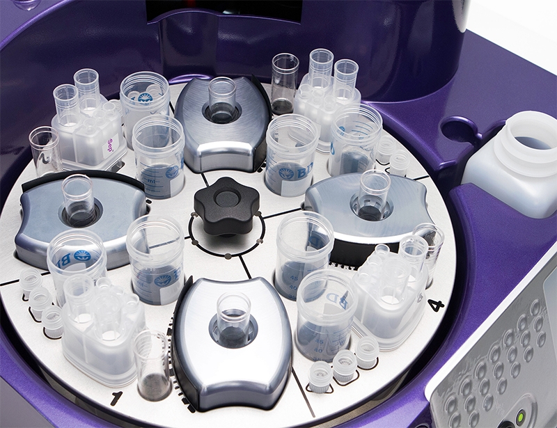
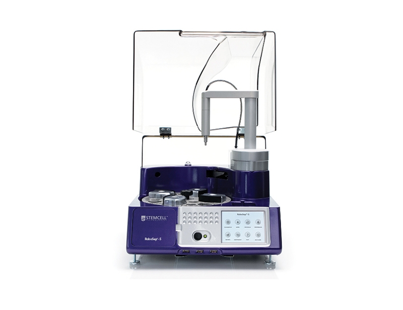
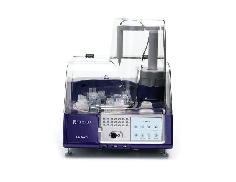
RoboSep™-S

RoboSep™-S - The Fully Automated Cell Separator
概要
RoboSep™-S is the next-generation RoboSep™ instrument for fully automated cell separation. Using EasySep™ technology, RoboSep™-S performs all steps necessary to magnetically label and separate virtually any cell type by positive or negative selection. RoboSep™-S is designed to minimize sample handling, eliminate cross-contamination, and reduce “hands-on” time.

To view cell isolation kits for use with RoboSep™-S, visit our Cell Isolation Products page.

Leasing options and warranty coverage are available. Please contact us for further information.

For more information about Instrument Services including additional service packages and software please see our instrumentation overview.                                
Contains
• RoboSep™-S Base Unit (Catalog #21001)
• "The Big Easy" EasySep™ Magnets (Catalog #18001)
• RoboSep™ Service Rack (Catalog #20101)
• RoboSep™ Tube Kits (Catalog #20155)
• USB Flash Drive
• RoboSep™ User Reference Manual (Catalog #29792)
• RoboSep™ Quick Start Guide (Catalog #28943)
• 1-Year Warranty (Catalog #21200)
Species
Human, Mouse, Rat, Non-Human Primate, Other
Application
Cell Isolation
Brand
RoboSep
Area of Interest
Chimerism, HLA, Immunology, Stem Cell Biology
数据及文献
Publications (63)

Leukemia 2019 mar

Selective targeting of multiple myeloma by B cell maturation antigen (BCMA)-specific central memory CD8+ cytotoxic T lymphocytes: immunotherapeutic application in vaccination and adoptive immunotherapy. 

J. Bae et al.                                    


To expand the breadth and extent of current multiple myeloma (MM)-specific immunotherapy, we have identified various antigens on CD138+ tumor cells from newly diagnosed MM patients (n = 616) and confirmed B-cell maturation antigen (BCMA) as a key myeloma-associated antigen. The aim of this study is to target the BCMA, which promotes MM cell growth and survival, by generating BCMA-specific memory CD8+ CTL that mediate effective and long-lasting immunity against MM. Here we report the identification of novel engineered peptides specific to BCMA, BCMA72-80 (YLMFLLRKI), and BCMA54-62 (YILWTCLGL), which display improved affinity/stability to HLA-A2 compared to their native peptides and induce highly functional BCMA-specific CTL with increased activation (CD38, CD69) and co-stimulatory (CD40L, OX40, GITR) molecule expression. Importantly, the heteroclitic BCMA72-80 specific CTL demonstrated poly-functional Th1-specific immune activities [IFN-gamma/IL-2/TNF-alpha production, proliferation, cytotoxicity] against MM, which were correlated with expansion of Tetramer+ and memory CD8+ CTL. Additionally, heteroclitic BCMA72-80 specific CTL treated with anti-OX40 (immune agonist) or anti-LAG-3 (checkpoint inhibitor) display increased immune function, mainly by central memory CTL. These results provide the framework for clinical application of heteroclitic BCMA72-80 peptide, alone and in combination with anti-LAG3 and/or anti-OX40 therapy, in vaccination and/or adoptive immunotherapeutic strategies to generate long-lasting anti-tumor immunity in patients with MM or other BCMA expressing tumors.


Nature metabolism 2019 jul

Metabolic plasticity of HIV-specific CD8+ T cells is associated with enhanced antiviral potential and natural control of HIV-1 infection. 

M. Angin et al.                                    

Spontaneous control of human immunodeficiency virus (HIV) is generally associated with an enhanced capacity of CD8+ T cells to eliminate infected CD4+ T cells, but the molecular characteristics of these highly functional CD8+ T cells are largely unknown. In the present study, using single-cell analysis, it was shown that HIV-specific, central memory CD8+ T cells from spontaneous HIV controllers (HICs) and antiretrovirally treated non-controllers have opposing transcriptomic profiles. Genes linked to effector functions and survival are upregulated in cells from HICs. In contrast, genes associated with activation, exhaustion and glycolysis are upregulated in cells from non-controllers. It was shown that HIV-specific CD8+ T cells from non-controllers are largely glucose dependent, whereas those from HICs have more diverse metabolic resources that enhance both their survival potential and their capacity to develop anti-HIV effector functions. The functional efficiency of the HIV-specific CD8+ T cell response in HICs is thus engraved in their memory population and related to their metabolic programme. Metabolic reprogramming in vitro through interleukin-15 treatment abrogated the glucose dependency and enhanced the antiviral potency of HIV-specific CD8+ T cells from non-controllers.


Immunity, inflammation and disease  2019

Complexin 2 regulates secretion of immunoglobulin in antibody-secreting cells. 

E. Tsuru et al. 

INTRODUCTION Complexins (CPLXs), initially identified in neuronal presynaptic terminals, are cytoplasmic proteins that interact with the soluble N-ethylmaleimide-sensitive factor attachment protein receptors (SNARE) complex to regulate the fusion of vesicles to the plasma membrane. Although much is known about CPLX function in neuronal synaptic vesicle exocytosis, their distribution and role in immune cells are still unclear. In this study, we investigated CPLX2 knockout (KO) mice to reveal the role of CPLXs in exocytosis of lymphocytes. METHODS We examined the expression of CPLXs and SNAREs in lymphocytes. To study the effect of CPLXs on the immune system in vivo, we analyzed the immune phenotype of CPLX2 KO mice. Furthermore, antibodies secretion from the peritoneal cavity, spleen, and bone marrow cells of wild-type (WT) and CPLX2 KO mice were determined. RESULTS CPLX2 was detected in B cells but not in T cells, while other CPLXs and SNAREs were expressed at a similar level in both B and T cells. To clarify the function of CPLX2 in B lymphocytes, serum concentrations of immunoglobulin G (IgG), IgA, IgM, and IgE were measured in WT and CPLX2 KO mice using enzyme-linked immunosorbent assay. The level of IgM, which mainly consists of natural antibodies, was higher in KO mice than that in WT mice, while the levels of other antibodies were similar in both types of mice. Additionally, we found that spontaneous secretion of IgM and IgG1 was enhanced from the splenic antibody-secreting cells (ASCs) of CPLX2 KO mice. CONCLUSION Our data suggest that CPLX2 inhibits spontaneous secretion of IgM and IgG1 from splenic ASCs. This study provides new insight into the mechanism of antibody secretion of ASCs.


The American journal of surgical pathology 2018 JUL

PD-1 Expression in Chronic Lymphocytic Leukemia/Small Lymphocytic Lymphoma (CLL/SLL) and Large B-cell Richter Transformation (DLBCL-RT): A Characteristic Feature of DLBCL-RT and Potential Surrogate Marker for Clonal Relatedness.

R. He et al.                                    

Chronic lymphocytic leukemia/small lymphocytic lymphoma (CLL/SLL) is a low-grade B-cell neoplasm and ∼2{\%} to 9{\%} patients develop an aggressive lymphoma, most commonly diffuse large B-cell lymphoma (Richter transformation, DLBCL-RT). Programmed death-1 (PD-1) pathway plays a crucial role in tumor host immunity evasion and its blockade has emerged as an effective anti-cancer immunotherapy. PD-L1 and PD-1 expression has shown predictive value in anti-PD cancer immunotherapy; however, it has not been well documented in CLL/SLL and DLBCL-RT. We evaluated PD-1 and PD-L1 expression by immunohistochemistry in 39 CLL/SLL, 15 DLBCL-RT, and 26 other DLBCL. In CLL/SLL, neoplastic B-cell PD-1 expression was weak and restricted to prolymphocytes/paraimmunoblasts within proliferation centers (PCs) and accentuated PCs of all sizes. Neoplastic B-cell PD-1 expression was highly prevalent and demonstrated increased intensity in DLBCL-RT, but in contrast was only rarely seen in other DLBCL (12/15 vs. 1/26; P{\textless}0.0001). An excellent correlation (90{\%} concordance) was observed between neoplastic B-cell PD-1 immunohistochemistry positivity and molecularly defined CLL/SLL clonal relatedness in DLBCL-RT. PD-L1 expression was observed on the neoplastic B cells in rare DLBCL-RT and other DLBCL cases (1/15 vs. 1/26; P{\textgreater}0.05) as well as background histiocytes and dendritic cells. Overall survival of DLBCL-RT was significantly inferior to that of the other DLBCL (median, 16.9 vs. 106.1 mo; P=0.002). Our findings suggest a biological continuum from prolymphocytes/paraimmunoblasts in CLL/SLL PCs to the neoplastic B-cells in DLBCL-RT. The characteristic PD-1 expression in DLBCL-RT makes it a potential surrogate marker for determining clonal relatedness to CLL/SLL, which may have important prognostic and therapeutic implications.


Cancer genetics 2018 DEC

Assessing genome-wide copy number aberrations and copy-neutral loss-of-heterozygosity as best practice: An evidence-based review from the Cancer Genomics Consortium working group for plasma cell disorders.

T. J. Pugh et al.                                    

BACKGROUND Plasma cell neoplasms (PCNs) encompass a spectrum of disorders including monoclonal gammopathy of undetermined significance, smoldering myeloma, plasma cell myeloma, and plasma cell leukemia. Molecular subtypes have been defined by recurrent cytogenetic abnormalities and somatic mutations that are prognostic and predictive. Karyotype and fluorescence in situ hybridization (FISH) have historically been used to guide management; however, new technologies and markers raise the need to reassess current testing algorithms. METHODS We convened a panel of representatives from international clinical laboratories to capture current state-of-the-art testing from published reports and to put forward recommendations for cytogenomic testing of plasma cell neoplasms. We reviewed 65 papers applying FISH, chromosomal microarray (CMA), next-generation sequencing, and gene expression profiling for plasma cell neoplasm diagnosis and prognosis. We also performed a survey of our peers to capture current laboratory practice employed outside our working group. RESULTS Plasma cell enrichment is widely used prior to FISH testing, most commonly by magnetic bead selection. A variety of strategies for direct, short- and long-term cell culture are employed to ensure clonal representation for karyotyping. Testing of clinically-informative 1p/1q, del(13q) and del(17p) are common using karyotype, FISH and, increasingly, CMA testing. FISH for a variety of clinically-informative balanced IGH rearrangements is prevalent. Literature review found that CMA analysis can detect abnormalities in 85-100{\%} of patients with PCNs; more specifically, in 5-53{\%} (median 14{\%}) of cases otherwise normal by FISH and cytogenetics. CMA results in plasma cell neoplasms are usually complex, with alteration counts ranging from 1 to 74 (median 10-20), primarily affecting loci not covered by FISH testing. Emerging biomarkers include structural alterations of MYC as well as somatic mutations of KRAS, NRAS, BRAF, and TP53. Together, these may be measured in a comprehensive manner by a combination of newer technologies including CMA and next-generation sequencing (NGS). Our survey suggests most laboratories have, or are soon to have, clinical CMA platforms, with a desire to move to NGS assays in the future. CONCLUSION We present an overview of current practices in plasma cell neoplasm testing as well as an algorithm for integrated FISH and CMA testing to guide treatment of this disease.


Scientific reports  2017 JAN

Non-pathogenic tissue-resident CD8+ T cells uniquely accumulate in the brains of lupus-prone mice.

P. A. Morawski et al.                                    

Severe lupus often includes psychiatric and neurological sequelae, although the cellular contributors to CNS disease remain poorly defined. Using intravascular staining to discriminate tissue-localized from blood-borne cells, we find substantial accumulation of CD8+ T cells relative to other lymphocytes in brain tissue, which correlates with lupus disease and limited neuropathology. This is in contrast to all other affected organs, where infiltrating CD4+ cells are predominant. Brain-infiltrating CD8+ T cells represent an activated subset of those found in the periphery, having a resident-memory phenotype (CD69+CD122-PD1+CD44+CD62L-) and expressing adhesion molecules (VLA-4+LFA-1+) complementary to activated brain endothelium. Remarkably, infiltrating CD8+ T cells do not cause tissue damage in lupus-prone mice, as genetic ablation of these cells via $\beta$2 m deficiency does not reverse neuropathology, but exacerbates disease both in the brain and globally despite decreased serum IgG levels. Thus, lupus-associated inflammation disrupts the blood-brain barrier in a discriminating way biased in favor of non-pathogenic CD8+ T cells relative to other infiltrating leukocytes, perhaps preventing further tissue damage in such a sensitive organ.
View All Publications                    
Top