Mouse Recombinant MIP-1 beta (CCL4)
Macrophage inflammatory protein-1 beta
概要
Macrophage inflammatory protein-1 beta (MIP-1 beta), also known as CCL4, is a member of the CC family of chemokines and is most closely related to CCL3 (MIP-1 alpha). Cellular sources of MIP-1 beta include activated leukocytes (monocytes and T and B cells), brain endothelial cells, and smooth muscle cells (Lukacs et al.; Menten et al.). MIP-1 beta, MIP-1 alpha, and RANTES have been shown to be major HIV-suppressive factors, possibly through the interactions of these chemokines with the receptor CCR5 on CD4+ T cells, which is also a major receptor for HIV entry into CD4+ T cells (Cocchi et al.; Menten et al.). MIP-1 beta attracts a variety of immune cells to sites of microbial infection. In addition to its chemotactic functions, MIP-1 beta induces the release of proinflammatory cytokines, mast cell degranulation, and NK cell activation (Schall et al.). In mice, recruitment of regulatory T cells to B cells and antigen-presenting cells by MIP-1 beta plays a central role in the initiation of T cell and humoral responses, and the depletion of regulatory T cells or MIP-1 beta results in deregulated humoral responses and production of autoantibodies (Bystry et al.).
Subtype
Cytokines
Alternative Names
ACT-2, Immune activation protein 2, LAG-1, Lymphocyte activation gene 1 protein, MIP-1b, Protein H400, SCYA2, SCYA4, Small-inducible cytokine A4, T-cell activation protein 2
Cell Type
B Cells, Dendritic Cells, Mesenchymal Stem and Progenitor Cells, Monocytes, NK Cells, Other, T Cells, T Cells, CD4+, T Cells, CD8+
Area of Interest
Immunology, Stem Cell Biology
Molecular Weight
7.8 kDa
Purity
≥ 95%
技术资料
| Document Type | 产品名称 | Catalog # | Lot # | 语言 |
|---|---|---|---|---|
| Product Information Sheet | Mouse Recombinant MIP-1 beta (CCL4) | 78091, 78091.1 | All | English |
| Safety Data Sheet | Mouse Recombinant MIP-1 beta (CCL4) | 78091, 78091.1 | All | English |
数据及文献
Data

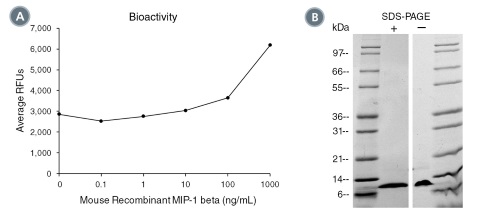
(A) The biological activity of Mouse Recombinant MIP-1 beta (CCL4) was tested by its ability to induce chemotaxis of THP-1 cells. Cell migration was measured after 45 min using a fluorometric assay method. Increase in migration over basal level was seen starting at 100 ng/mL. (B) 1 μg of Mouse Recombinant MIP-1 beta (CCL4) was resolved with SDS-PAGE under reducing (+) and non-reducing (-) conditions and visualized by Coomassie Blue staining. Mouse Recombinant MIP-1 beta (CCL4) has a predicted molecular mass of 7.8 kDa.


