EasySep™ Human Plasmacytoid DC Enrichment Kit
Immunomagnetic negative selection cell isolation kit
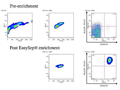
概要
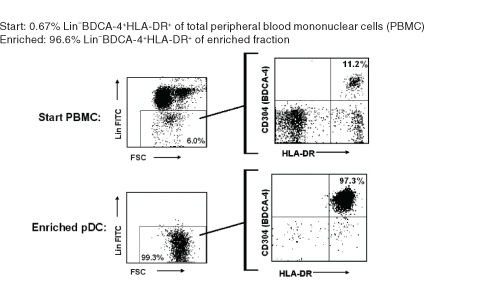
The EasySep™ Human Plasmacytoid DC Enrichment Kit is designed to isolate plasmacytoid dendritic cells (pDC) from fresh peripheral blood mononuclear cells by negative selection. Unwanted cells are targeted for removal with Tetrameric Antibody Complexes recognizing non-pDCs and dextran-coated magnetic particles. The labeled cells are separated using the EasySep™ magnet without the use of columns. Desired cells are poured off into a new tube.
• Fast, easy-to-use and column-free
• Up to 97% purity
• Untouched, viable cells
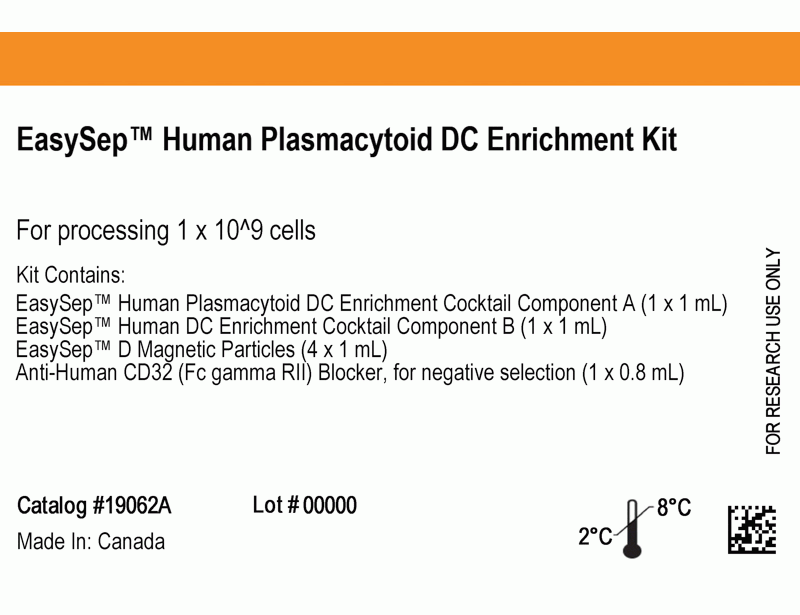
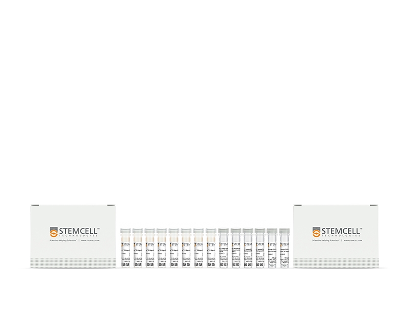
- EasySep™ Human Plasmacytoid DC Enrichment Kit (Catalog #19062)
- EasySep™ Human Plasmacytoid DC Enrichment Cocktail Component A, 2 x 1 mL
- EasySep™ Human DC Enrichment Cocktail Component B, 2 x 1 mL
- EasySep™ D Magnetic Particles, 8 x 1 mL
- Anti-Human CD32 (Fc gamma RII) Blocker, 2 x 0.8 mL
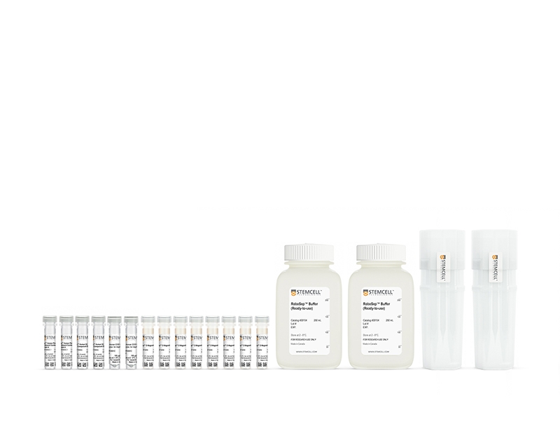
- RoboSep™ Human Plasmacytoid DC Enrichment Kit with Filter Tips (Catalog #19062RF)
- EasySep™ Human Plasmacytoid DC Enrichment Cocktail Component A, 2 x 1 mL
- EasySep™ Human DC Enrichment Cocktail Component B, 2 x 1 mL
- EasySep™ D Magnetic Particles, 8 x 1 mL
- Anti-Human CD32 (Fc gamma RII) Blocker, 2 x 0.8 mL
- RoboSep™ Buffer (Catalog #20104) x 2
- RoboSep™ Filter Tips (Catalog #20125) x 2
• EasySep™ Magnet (Catalog #18000)
• “The Big Easy” EasySep™ Magnet (Catalog #18001)
• EasyPlate™ EasySep™ Magnet (Catalog 18102)
• Easy 50 EasySep™ Magnet (Catalog #18002)
• RoboSep™-S (Catalog #21000)
数据及文献
Publications (12)
Journal of immunology (Baltimore, Md. : 1950) 2018 NOV
Differential and Overlapping Immune Programs Regulated by IRF3 and IRF5 in Plasmacytoid Dendritic Cells.
K. T. Chow et al.
Abstract
We examined the signaling pathways and cell type-specific responses of IFN regulatory factor (IRF) 5, an immune-regulatory transcription factor. We show that the protein kinases IKK$\alpha$, IKK$\beta$, IKK$\epsilon$, and TANK-binding kinase 1 each confer IRF5 phosphorylation/dimerization, thus extending the family of IRF5 activator kinases. Among primary human immune cell subsets, we found that IRF5 is most abundant in plasmacytoid dendritic cells (pDCs). Flow cytometric cell imaging revealed that IRF5 is specifically activated by endosomal TLR signaling. Comparative analyses revealed that IRF3 is activated in pDCs uniquely through RIG-I-like receptor (RLR) signaling. Transcriptomic analyses of pDCs show that the partitioning of TLR7/IRF5 and RLR/IRF3 pathways confers differential gene expression and immune cytokine production in pDCs, linking IRF5 with immune regulatory and proinflammatory gene expression. Thus, TLR7/IRF5 and RLR-IRF3 partitioning serves to polarize pDC response outcome. Strategies to differentially engage IRF signaling pathways should be considered in the design of immunotherapeutic approaches to modulate or polarize the immune response for specific outcome.
Nature immunology 2018 JAN
Diversification of human plasmacytoid predendritic cells in response to a single stimulus.
Alculumbre SG et al.
Abstract
Innate immune cells adjust to microbial and inflammatory stimuli through a process termed environmental plasticity, which links a given individual stimulus to a unique activated state. Here, we report that activation of human plasmacytoid predendritic cells (pDCs) with a single microbial or cytokine stimulus triggers cell diversification into three stable subpopulations (P1-P3). P1-pDCs (PD-L1+CD80-) displayed a plasmacytoid morphology and specialization for type I interferon production. P3-pDCs (PD-L1-CD80+) adopted a dendritic morphology and adaptive immune functions. P2-pDCs (PD-L1+CD80+) displayed both innate and adaptive functions. Each subpopulation expressed a specific coding- and long-noncoding-RNA signature and was stable after secondary stimulation. P1-pDCs were detected in samples from patients with lupus or psoriasis. pDC diversification was independent of cell divisions or preexisting heterogeneity within steady-state pDCs but was controlled by a TNF autocrine and/or paracrine communication loop. Our findings reveal a novel mechanism for diversity and division of labor in innate immune cells.
The Journal of allergy and clinical immunology 2017 SEP
Enhanced plasmacytoid dendritic cell antiviral responses after omalizumab.
Gill MA et al.
Abstract
BACKGROUND Atopy and viral respiratory tract infections synergistically promote asthma exacerbations. IgE cross-linking inhibits critical virus-induced IFN-α responses of plasmacytoid dendritic cells (pDCs), which can be deficient in patients with allergic asthma. OBJECTIVE We sought to determine whether reducing IgE levels in vivo with omalizumab treatment increases pDC antiviral IFN-α responses in inner-city children with asthma. METHODS PBMCs and pDCs isolated from children with exacerbation-prone asthma before and during omalizumab treatment were stimulated ex vivo with rhinovirus and influenza in the presence or absence of IgE cross-linking. IFN-α levels were measured in supernatants, and mRNA expression of IFN-α pathway genes was determined by using quantitative RT-PCR (qRT-PCR) in cell pellets. FcεRIα protein levels and mRNA expression were measured in unstimulated cells by using flow cytometry and qRT-PCR, respectively. Changes in these outcomes and associations with clinical outcomes were analyzed, and statistical modeling was used to identify risk factors for asthma exacerbations. RESULTS Omalizumab treatment increased rhinovirus- and influenza-induced PBMC and rhinovirus-induced pDC IFN-α responses in the presence of IgE cross-linking and reduced pDC surface FcεRIα expression. Omalizumab-induced reductions in pDC FcεRIα levels were significantly associated with a lower asthma exacerbation rate during the outcome period and correlated with increases in PBMC IFN-α responses. PBMC FcεRIα mRNA expression measured on study entry significantly improved an existing model of exacerbation prediction. CONCLUSIONS These findings indicate that omalizumab treatment augments pDC IFN-α responses and attenuates pDC FcεRIα protein expression and provide evidence that these effects are related. These results support a potential mechanism underlying clinical observations that allergic sensitization is associated with increased susceptibility to virus-induced asthma exacerbations.
Blood 2017
NOX5 and p22phox are 2 novel regulators of human monocytic differentiation into dendritic cells.
Marzaioli V et al.
Abstract
Dendritic cells (DCs) are a heterogeneous population of professional antigen-presenting cells and are key cells of the immune system, acquiring different phenotypes in accordance with their localization during the immune response. A subset of inflammatory DCs is derived from circulating monocytes (Mo) and has a key role in inflammation and infection. The pathways controlling Mo-DC differentiation are not fully understood. Our objective was to investigate the possible role of nicotinamide adenine dinucleotide phosphate reduced form oxidases (NOXs) in Mo-DC differentiation. In this study, we revealed that Mo-DC differentiation was inhibited by NOX inhibitors and reactive oxygen species scavengers. We show that the Mo-DC differentiation was dependent on p22phox, and not on gp91phox/NOX2, as shown by the reduced Mo-DC differentiation observed in chronic granulomatous disease patients lacking p22phox. Moreover, we revealed that NOX5 expression was strongly increased during Mo-DC differentiation, but not during Mo-macrophage differentiation. NOX5 was expressed in circulating myeloid DC, and at a lower level in plasmacytoid DC. Interestingly, NOX5 was localized at the outer membrane of the mitochondria and interacted with p22phox in Mo-DC. Selective inhibitors and small interfering RNAs for NOX5 indicated that NOX5 controlled Mo-DC differentiation by regulating the JAK/STAT/MAPK and NFκB pathways. These data demonstrate that the NOX5-p22phox complex drives Mo-DC differentiation, and thus could be critical for immunity and inflammation.
PLoS pathogens 2016 FEB
HMGB1 Is Involved in IFN-α Production and TRAIL Expression by HIV-1-Exposed Plasmacytoid Dendritic Cells: Impact of the Crosstalk with NK Cells.
Saï et al.
Abstract
Plasmacytoid dendritic cells (pDCs) are innate sensors of viral infections and important mediators of antiviral innate immunity through their ability to produce large amounts of IFN-α. Moreover, Toll-like receptor 7 (TLR7) and 9 (TLR9) ligands, such as HIV and CpG respectively, turn pDCs into TRAIL-expressing killer pDCs able to lyse HIV-infected CD4+ T cells. NK cells can regulate antiviral immunity by modulating pDC functions, and pDC production of IFN-α as well as cell-cell contact is required to promote NK cell functions. Impaired pDC-NK cell crosstalk was reported in the setting of HIV-1 infection, but the impact of HIV-1 on TRAIL expression and innate antiviral immunity during this crosstalk is unknown. Here, we report that low concentrations of CCR5-tropic HIV-1Ba-L promote the release of pro-inflammatory cytokines such as IFN-α, TNF-α, IFN-γ and IL-12, and CCR5-interacting chemokines (MIP-1α and MIP-1β) in NK-pDCs co-cultures. At high HIV-1BaL concentrations, the addition of NK cells did not promote the release of these mediators, suggesting that once efficiently triggered by the virus, pDCs could not integrate new activating signals delivered by NK cells. However, high HIV-1BaL concentrations were required to trigger IFN-α-mediated TRAIL expression at the surface of both pDCs and NK cells during their crosstalk. Interestingly, we identified the alarmin HMGB1, released at pDC-NK cell synapse, as an essential trigger for the secretion of IFN-α and IFN-related soluble mediators during the interplay of HIV-1 exposed pDCs with NK cells. Moreover, HMGB1 was found crucial for mTRAIL translocation to the plasma membrane of both pDCs and NK cells during their crosstalk following pDC exposure to HIV-1. Data from serum analyses of circulating HMGB1, HMGB1-specific antibodies, sTRAIL and IP-10 in a cohort of 67 HIV-1+ patients argue for the in vivo relevance of these observations. Altogether, these findings identify HMGB1 as a trigger for IFN-α-mediated TRAIL expression at the surface of pDCs and NK cells, and they suggest a novel mechanism of innate control of HIV-1 infection.
Oncotarget 2015 OCT
TRAIL-mediated killing of acute lymphoblastic leukemia by plasmacytoid dendritic cell-activated natural killer cells.
Lelaidier M et al.
Abstract
Acute lymphoblastic leukemia (ALL) still frequently recurs after hematopoietic stem cell transplantation (HSCT), underscoring the need to improve the graft-versus-leukemia (GvL) effect. Natural killer (NK) cells reconstitute in the first months following HSCT when leukemia burden is at its lowest, but ALL cells have been shown to be resistant to NK cell-mediated killing. We show here that this resistance is overcome by NK cell stimulation with TLR-9-activated plasmacytoid dendritic cells (pDCs). NK cell priming with activated pDCs resulted in TRAIL and CD69 up-regulation on NK cells and IFN-γ production. NK cell activation was dependent on IFN-α produced by pDCs, but was not reproduced by IFN-α alone. ALL killing was further enhanced by inhibition of KIR engagement. We showed that ALL lysis was mainly mediated by TRAIL engagement, while the release of cytolytic granules was involved when ALL expressed NK cell activating receptor ligands. Finally, adoptive transfers of activated-pDCs in ALL-bearing humanized mice delayed the leukemia onset and cure 30% of mice. Our data therefore demonstrate that TLR-9 activated pDCs are a powerful tool to overcome ALL resistance to NK cell-mediated killing and to reinforce the GvL effect of HSCT. These results open new therapeutic avenues to prevent relapse in children with ALL.
View All Publications