
EasySep™ Human NK Cell Enrichment Kit
Immunomagnetic negative selection cell isolation kit
概要
The EasySep™ Human NK Cell Enrichment Kit is designed to isolate NK cells from fresh or previously frozen peripheral blood mononuclear cells by negative selection. Unwanted cells are targeted for removal with Tetrameric Antibody Complexes recognizing non-NK cells and dextran-coated magnetic particles. The labeled cells are separated using an EasySep™ magnet without the use of columns. Desired cells are poured off into a new tube.
For even faster cell isolations, we recommend the new EasySep™ Human NK Cell Isolation Kit (17955) which isolates cells in just 8 minutes.
For even faster cell isolations, we recommend the new EasySep™ Human NK Cell Isolation Kit (17955) which isolates cells in just 8 minutes.
Advantages
• Fast, easy-to-use and column-free
• Up to 95% purity
• Untouched, viable cells
• Up to 95% purity
• Untouched, viable cells
Components
- EasySep™ Human NK Cell Enrichment Kit (Catalog #19055)
- EasySep™ Human NK Cell Enrichment Cocktail, 1 mL
- EasySep™ D Magnetic Particles, 2 x 1 mL
- RoboSep™ Human NK Cell Enrichment Kit with Filter Tips (Catalog #19055RF)
- EasySep™ Human NK Cell Enrichment Cocktail, 1 mL
- EasySep™ D Magnetic Particles, 2 x 1 mL
- RoboSep™ Buffer (Catalog #20104)
- RoboSep™ Filter Tips (Catalog #20125)
Magnet Compatibility
• EasySep™ Magnet (Catalog #18000)
• “The Big Easy” EasySep™ Magnet (Catalog #18001)
• Easy 50 EasySep™ Magnet (Catalog #18002)
• EasyPlate™ EasySep™ Magnet (Catalog 18102)
• EasyEights™ EasySep™ Magnet (Catalog #18103)
• RoboSep™-S (Catalog #21000)
Subtype
Cell Isolation Kits
Cell Type
NK Cells
Species
Human
Sample Source
PBMC
Selection Method
Negative
Application
Cell Isolation
Brand
EasySep, RoboSep
Area of Interest
Immunology
技术资料
| Document Type | 产品名称 | Catalog # | Lot # | 语言 |
|---|---|---|---|---|
| Product Information Sheet | EasySep™ Human NK Cell Enrichment Kit | 19055 | All | English |
| Product Information Sheet | RoboSep™ Human NK Cell Enrichment Kit with Filter Tips | 19055RF | All | English |
| Safety Data Sheet | EasySep™ Human NK Cell Enrichment Kit | 19055 | All | English |
| Safety Data Sheet 1 | RoboSep™ Human NK Cell Enrichment Kit with Filter Tips | 19055RF | All | English |
| Safety Data Sheet 2 | RoboSep™ Human NK Cell Enrichment Kit with Filter Tips | 19055RF | All | English |
数据及文献
Data
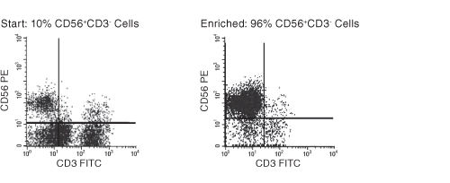
Figure 1. FACS Profile Results With EasySep™ Human NK Cell Enrichment Kit
The NK cell content of the enriched fraction varies, depending on the starting sample. Starting with previously frozen mononuclear cells containing more than 10% NK cells, the NK cell content of the enriched fraction typically ranges from 73% - 95%. Purities may be lower when starting with samples containing less than 10% NK cells.
Publications (35)
Cell stem cell 2020 jun
Metabolic Reprograming via Deletion of CISH in Human iPSC-Derived NK Cells Promotes In Vivo Persistence and Enhances Anti-tumor Activity.
Abstract
Abstract
Cytokine-inducible SH2-containing protein (CIS; encoded by the gene CISH) is a key negative regulator of interleukin-15 (IL-15) signaling in natural killer (NK) cells. Here, we develop human CISH-knockout (CISH-/-) NK cells using an induced pluripotent stem cell-derived NK cell (iPSC-NK cell) platform. CISH-/- iPSC-NK cells demonstrate increased IL-15-mediated JAK-STAT signaling activity. Consequently, CISH-/- iPSC-NK cells exhibit improved expansion and increased cytotoxic activity against multiple tumor cell lines when maintained at low cytokine concentrations. CISH-/- iPSC-NK cells display significantly increased in vivo persistence and inhibition of tumor progression in a leukemia xenograft model. Mechanistically, CISH-/- iPSC-NK cells display improved metabolic fitness characterized by increased basal glycolysis, glycolytic capacity, maximal mitochondrial respiration, ATP-linked respiration, and spare respiration capacity mediated by mammalian target of rapamycin (mTOR) signaling that directly contributes to enhanced NK cell function. Together, these studies demonstrate that CIS plays a key role to regulate human NK cell metabolic activity and thereby modulate anti-tumor activity.
Scientific reports 2020 feb
The anti-inflammatory potential of cefazolin as common gamma chain cytokine inhibitor.
Abstract
Abstract
A continuing quest for specific inhibitors of proinflammatory cytokines brings promise for effective therapies designed for inflammatory and autoimmune disorders. Cefazolin, a safe, first-generation cephalosporin antibiotic, has been recently shown to specifically interact with interleukin 15 (IL-15) receptor subunit $\alpha$ (IL-15R$\alpha$) and to inhibit IL-15-dependent TNF-$\alpha$ and IL-17 synthesis. The aim of this study was to elucidate cefazolin activity against IL-2, IL-4, IL-15 and IL-21, i.e. four cytokines sharing the common cytokine receptor $\gamma$ chain ($\gamma$c). In silico, molecular docking unveiled two potential cefazolin binding sites within the IL-2/IL-15R$\beta$ subunit and two within the $\gamma$c subunit. In vitro, cefazolin decreased proliferation of PBMC (peripheral blood mononuclear cells) following IL-2, IL-4 and IL-15 stimulation, reduced production of IFN-$\gamma$, IL-17 and TNF-$\alpha$ in IL-2- and IL-15-treated PBMC and in IL-15 stimulated natural killer (NK) cells, attenuated IL-4-dependent expression of CD11c in monocyte-derived dendritic cells and suppressed phosphorylation of JAK3 in response to IL-2 and IL-15 in PBMC, to IL-4 in TF-1 (erythroleukemic cell line) and to IL-21 in NK-92 (NK cell line). The results of the study suggest that cefazolin may exert inhibitory activity against all of the $\gamma$c receptor-dependent cytokines, i.e. IL-2, IL-4, IL-7, IL-9, IL-15 and IL-21.
Nature chemical biology 2020 dec
Targeted glycan degradation potentiates the anticancer immune response in vivo.
Abstract
Abstract
Currently approved immune checkpoint inhibitor therapies targeting the PD-1 and CTLA-4 receptor pathways are powerful treatment options for certain cancers; however, most patients across cancer types still fail to respond. Consequently, there is interest in discovering and blocking alternative pathways that mediate immune suppression. One such mechanism is an upregulation of sialoglycans in malignancy, which has been recently shown to inhibit immune cell activation through multiple mechanisms and therefore represents a targetable glycoimmune checkpoint. Since these glycans are not canonically druggable, we designed an $\alpha$HER2 antibody-sialidase conjugate that potently and selectively strips diverse sialoglycans from breast cancer cells. In syngeneic breast cancer models, desialylation enhanced immune cell infiltration and activation and prolonged the survival of mice, an effect that was dependent on expression of the Siglec-E checkpoint receptor found on tumor-infiltrating myeloid cells. Thus, antibody-sialidase conjugates represent a promising modality for glycoimmune checkpoint therapy.
Pathogens (Basel, Switzerland) 2019 nov
Distinct Effects of Immunosuppressive Drugs on the Anti-Aspergillus Activity of Human Natural Killer Cells.
Abstract
Abstract
As the prognosis of invasive aspergillosis remains unacceptably poor in patients undergoing hematopoietic stem cell transplantation (HSCT), there is a growing interest in the adoptive transfer of antifungal effector cells, such as Natural Killer (NK) cells. Because immunosuppressive agents are required in most HSCT recipients, knowledge of the impact of these compounds on the antifungal activity of NK cells is a prerequisite for clinical trials. We, therefore, assessed the effect of methylprednisolone (mPRED), cyclosporin A (CsA) and mycophenolic acid (MPA) at different concentrations on proliferation, apoptosis/necrosis, and the direct and indirect anti-Aspergillus activity of human NK cells. Methylprednisolone decreased proliferation and increased apoptosis of NK cells in a significant manner. After seven days, a reduction of viable NK cells was seen for all three immunosuppressants, which was significant for MPA only. Cyclosporin A significantly inhibited the direct hyphal damage by NK cells in a dose-dependent manner. None of the immunosuppressive compounds had a major impact on the measured levels of interferon-$\gamma$, granulocyte-macrophage colony-stimulating factor and RANTES (regulated on activation, normal T cell expressed and secreted; CCL5). Our data demonstrate that commonly used immunosuppressive compounds have distinct effects on proliferation, viability and antifungal activity of human NK cells, which should be considered in designing studies on the use of NK cells for adoptive antifungal immunotherapy.
Cell 2019 may
A Site of Vulnerability on the Influenza Virus Hemagglutinin Head Domain Trimer Interface.
Abstract
Abstract
Here, we describe the discovery of a naturally occurring human antibody (Ab), FluA-20, that recognizes a new site of vulnerability on the hemagglutinin (HA) head domain and reacts with most influenza A viruses. Structural characterization of FluA-20 with H1 and H3 head domains revealed a novel epitope in the HA trimer interface, suggesting previously unrecognized dynamic features of the trimeric HA protein. The critical HA residues recognized by FluA-20 remain conserved across most subtypes of influenza A viruses, which explains the Ab's extraordinary breadth. The Ab rapidly disrupted the integrity of HA protein trimers, inhibited cell-to-cell spread of virus in culture, and protected mice against challenge with viruses of H1N1, H3N2, H5N1, or H7N9 subtypes when used as prophylaxis or therapy. The FluA-20 Ab has uncovered an exceedingly conserved protective determinant in the influenza HA head domain trimer interface that is an unexpected new target for anti-influenza therapeutics and vaccines.
Scientific reports 2019 feb
High percentages and activity of synovial fluid NK cells present in patients with advanced stage active Rheumatoid Arthritis.
Abstract
Abstract
Rheumatoid Arthritis (RA) causes chronic inflammation of joints. The cytokines TNFalpha and IFNgamma are central players in RA, however their source has not been fully elucidated. Natural Killer (NK) cells are best known for their role in elimination of viral-infected and transformed cells, and they secrete pro-inflammatory cytokines. NK cells are present in the synovial fluids (SFs) of RA patients and are considered to be important in bone destruction. However, the phenotype and function of NK cells in the SFs of patients with erosive deformative RA (DRA) versus non-deformative RA (NDRA) is poorly characterized. Here we characterize the NK cell populations present in the blood and SFs of DRA and NDRA patients. We demonstrate that a distinct population of activated synovial fluid NK (sfNK) cells constitutes a large proportion of immune cells found in the SFs of DRA patients. We discovered that although sfNK cells in both DRA and NDRA patients have similar phenotypes, they function differently. The DRA sfNK secrete more TNFalpha and IFNgamma upon exposure to IL-2 and IL-15. Consequently, we suggest that sfNK cells may be a marker for more severely destructive RA disease.