RosetteSep™ Human Mesenchymal Stem Cell Enrichment Cocktail
Immunodensity negative selection cocktail
概要
The RosetteSep™ Human Mesenchymal Stem Cell Enrichment Cocktail is designed to isolate mesenchymal stem cells from fresh bone marrow by negative selection. Unwanted cells are targeted for removal with Tetrameric Antibody Complexes recognizing CD3, CD14, CD19, CD38, CD66b and glycophorin A on red blood cells (RBCs). When centrifuged over a buoyant density medium such as Lymphoprep™ (Catalog #07801), the unwanted cells pellet along with the RBCs. The purified mesenchymal stem cells are present as a highly enriched population at the interface between the plasma and the buoyant density medium.
Advantages
• Fast and easy-to-use
• Requires no special equipment or training
• Untouched, viable cells
• Requires no special equipment or training
• Untouched, viable cells
Components
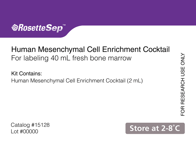
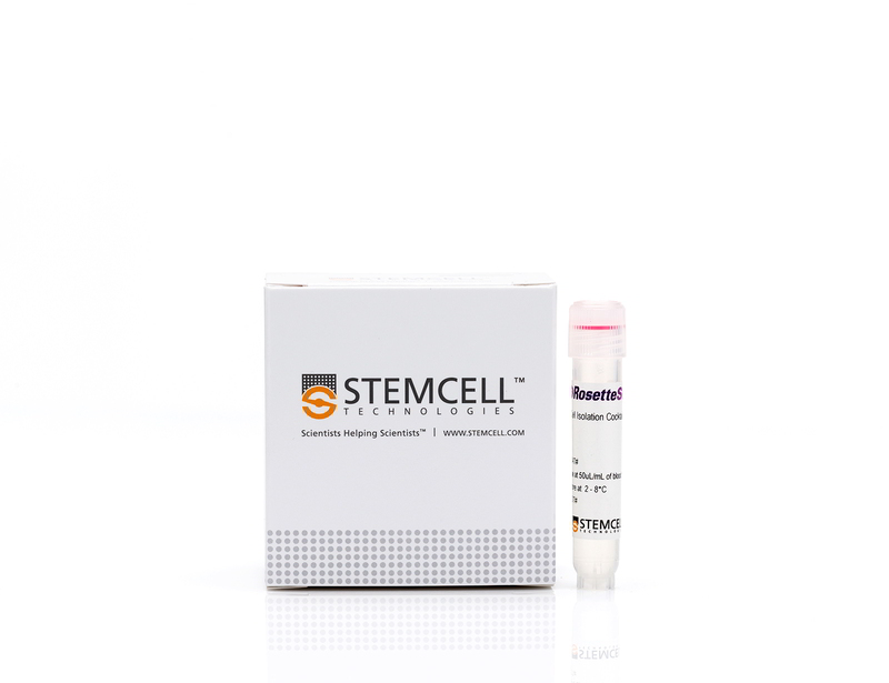
- RosetteSep™ Human Mesenchymal Stem Cell Enrichment Cocktail (Catalog #15128)
- RosetteSep™ Human Mesenchymal Stem Cell Enrichment Cocktail, 2 mL
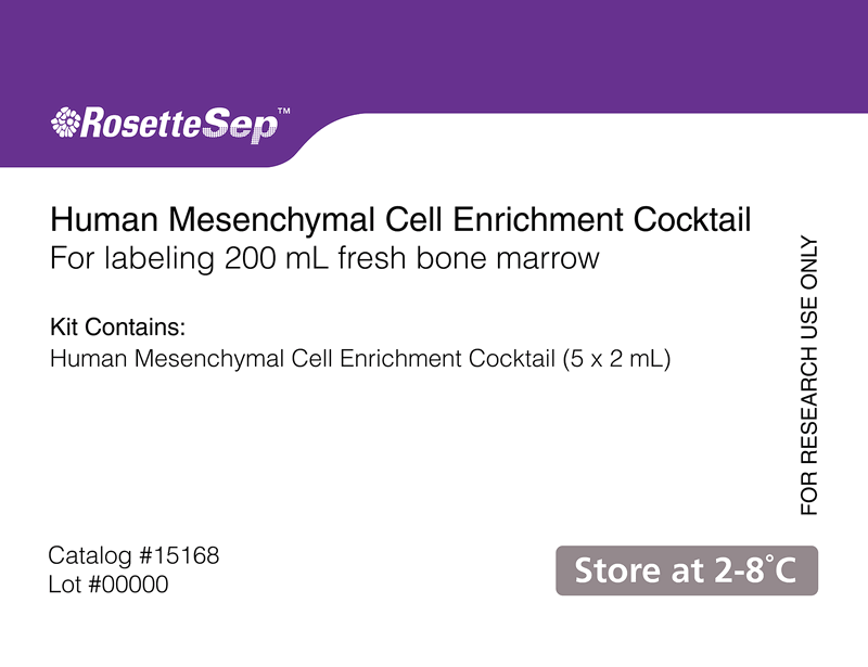
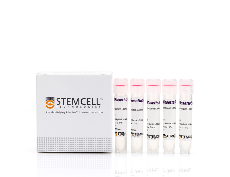
- RosetteSep™ Human Mesenchymal Stem Cell Enrichment Cocktail (Catalog #15168)
- RosetteSep™ Human Mesenchymal Stem Cell Enrichment Cocktail, 5 x 2 mL
Subtype
Cell Isolation Kits
Cell Type
Mesenchymal Stem and Progenitor Cells
Species
Human
Sample Source
Bone Marrow
Selection Method
Negative
Application
Cell Isolation
Brand
RosetteSep
Area of Interest
Stem Cell Biology
技术资料
| Document Type | 产品名称 | Catalog # | Lot # | 语言 |
|---|---|---|---|---|
| Product Information Sheet | RosetteSep™ Human Mesenchymal Stem Cell Enrichment Cocktail | 15128, 15168 | All | English |
| Safety Data Sheet | RosetteSep™ Human Mesenchymal Stem Cell Enrichment Cocktail | 15128 | All | English |
| Safety Data Sheet | RosetteSep™ Human Mesenchymal Stem Cell Enrichment Cocktail | 15168 | All | English |
数据及文献
Publications (7)
Clinical cancer research : an official journal of the American Association for Cancer Research 2019 aug
Auranofin Protects Intestine against Radiation Injury by Modulating p53/p21 Pathway and Radiosensitizes Human Colon Tumor.
Abstract
Abstract
PURPOSE The radiosensitivity of the normal intestinal epithelium is the major limiting factor for definitive radiotherapy against abdominal malignancies. Radiosensitizers, which can be used without augmenting radiation toxicity to normal tissue, are still an unmet need. Inhibition of proteosomal degradation is being developed as a major therapeutic strategy for anticancer therapy as cancer cells are more susceptible to proteasomal inhibition-induced cytotoxicity compared with normal cells. Auranofin, a gold-containing antirheumatoid drug, blocks proteosomal degradation by inhibiting deubiquitinase inhibitors. In this study, we have examined whether auranofin selectively radiosensitizes colon tumors without promoting radiation toxicity in normal intestine. EXPERIMENTAL DESIGN The effect of auranofin (10 mg/kg i.p.) on the radiation response of subcutaneous CT26 colon tumors and the normal gastrointestinal epithelium was determined using a mouse model of abdominal radiation. The effect of auranofin was also examined in a paired human colonic organoid system using malignant and nonmalignant tissues from the same patient. RESULTS Both in the mouse model of intestinal injury and in the human nonmalignant colon organoid culture, auranofin pretreatment prevented radiation toxicity and improved survival with the activation of p53/p21-mediated reversible cell-cycle arrest. However, in a mouse model of abdominal tumor and in human malignant colonic organoids, auranofin inhibited malignant tissue growth with inhibition of proteosomal degradation, induction of endoplasmic reticulum stress/unfolded protein response, and apoptosis. CONCLUSIONS Our data suggest that auranofin is a potential candidate to be considered as a combination therapy with radiation to improve therapeutic efficacy against abdominal malignancies.
Cell biology international 2012 JUL
New approach to isolate mesenchymal stem cell (MSC) from human umbilical cord blood.
Abstract
Abstract
HUCB (human umbilical cord blood) has been frequently used in clinical allogeneic HSC (haemopoietic stem cell) transplant. However, HUCB is poorly recognized as a rich source of MSC (mesenchymal stem cell). The aim of this study has been to establish a new method for isolating large number of MSC from HUCB to recognize it as a good source of MSC. HUCB samples were collected from women following their elective caesarean section. The new method (Clot Spot method) was carried out by explanting HUCB samples in mesencult complete medium and maintained in 37°C, in a 5% CO2 and air incubator. MSC presence was established by quantitative and qualitative immunophenotyping of cells and using FITC attached to MSC phenotypic markers (CD29, CD73, CD44 and CD105). Haematopoietic antibodies (CD34 and CD45) were used as negative control. MSC differentiation was examined in neurogenic and adipogenic media. Immunocytochemistry was carried out for the embryonic markers: SOX2 (sex determining region Y-box 2), OLIG-4 (oligodendrocyte-4) and FABP-4 (fatty acid binding protein-4). The new method was compared with the conventional Rosset Sep method. MSC cultures using the Clot Spot method showed 3-fold increase in proliferation rate compared with conventional method. Also, the cells showed high expression of MSC markers CD29, CD73, CD44 and CD105, but lacked the expression of specific HSC markers (CD34 and CD45). The isolated MSC showed some differentiation by expressing the neurogenic (SOX2 and Olig4) and adipogenic (FABP-4) markers respectively. In conclusion, HUCB is a good source of MSC using this new technique.
Bone 2008 DEC
Gene expression analysis in osteoblastic differentiation from peripheral blood mesenchymal stem cells.
Abstract
Abstract
MSCs are known to have an extensive proliferative potential and ability to differentiate in various cell types. Osteoblastic differentiation from mesenchymal progenitor cells is an important step of bone formation, though the pattern of gene expression during differentiation is not yet well understood. Here, to investigate the possibility to obtain a model for in vitro bone differentiation using mesenchymal stem cells (hMSCs) from human subjects non-invasively, we developed a method to obtain hMSCs-like cells from peripheral blood by a two step method that included an enrichment of mononuclear cells followed by depletion of unwanted cells. Using these cells, we analyzed the expression of transcription factor genes (runt-related transcription factor 2 (RUNX2) and osterix (SP7)) and bone related genes (osteopontin (SPP1), osteonectin (SPARC) and collagen, type I, alpha 1 (COLIA1)) during osteoblastic differentiation. Our results demonstrated that hMSCs can be obtained from peripheral blood and that they are able to generate CFU-F and to differentiate in osteoblast and adipocyte; in this study, we also identified a possible gene expression timing during osteoblastic differentiation that provided a powerful tool to study bone physiology.
Blood 2007 JUL
Identification of functional endothelial progenitor cells suitable for the treatment of ischemic tissue using human umbilical cord blood.
Abstract
Abstract
Umbilical cord blood (UCB) has been used as a potential source of various kinds of stem cells, including hematopoietic stem cells, mesenchymal stem cells, and endothelial progenitor cells (EPCs), for a variety of cell therapies. Recently, EPCs were introduced for restoring vascularization in ischemic tissues. An appropriate procedure for isolating EPCs from UCB is a key issue for improving therapeutic efficacy and eliminating the unexpected expansion of nonessential cells. Here we report a novel method for isolating EPCs from UCB by a combination of negative immunoselection and cell culture techniques. In addition, we divided EPCs into 2 subpopulations according to the aldehyde dehydrogenase (ALDH) activity. We found that EPCs with low ALDH activity (Alde-Low) possess a greater ability to proliferate and migrate compared to those with high ALDH activity (Alde-High). Moreover, hypoxia-inducible factor proteins are up-regulated and VEGF, CXCR4, and GLUT-1 mRNAs are increased in Alde-Low EPCs under hypoxic conditions, while the response was not significant in Alde-High EPCs. In fact, the introduction of Alde-Low EPCs significantly reduced tissue damage in ischemia in a mouse flap model. Thus, the introduction of Alde-Low EPCs may be a potential strategy for inducing rapid neovascularization and subsequent regeneration of ischemic tissues.
Journal of lipid research 2007 AUG
Direct evidence of lipid translocation between adipocytes and prostate cancer cells with imaging FTIR microspectroscopy.
Abstract
Abstract
Various epidemiological studies show a positive correlation between high intake of dietary FAs and metastatic prostate cancer (CaP). Moreover, CaP metastasizes to the bone marrow, which harbors a rich source of lipids stored within adipocytes. Here, we use Fourier transform infrared (FTIR) microspectroscopy to study adipocyte biochemistry and to demonstrate that PC-3 cells uptake isotopically labeled FA [deuterated palmitic acid (D(31)-PA)] from an adipocyte. Using this vibrational spectroscopic technique, we detected subcellular locations in a single adipocyte enriched with D(31)-PA using the upsilon(as+s)(C-D)(2+3) (D(31)-PA): upsilon(as+s)(C-H)(2+3) (lipid hydrocarbon) signal. In addition, larger adipocytes were found to consist of a higher percentage of D(31)-PA of the total lipid found within the adipocyte. Following background subtraction, the upsilon(as)(C-D)(2+3) signal illuminated starved PC-3 cells cocultured with D(31)-PA-loaded adipocytes, indicating translocation of the labeled FA. This study demonstrates lipid-specific translocation between adipocytes and tumor cells and the use of FTIR microspectroscopy to characterize various biomolecular features of a single adipocyte without the requirement for cell isolation and lipid extraction.
Experimental cell research 2005 JUL
Contribution of human bone marrow stem cells to individual skeletal myotubes followed by myogenic gene activation.
Abstract
Abstract
Much attention is focused on characterizing the contribution of bone marrow (BM)-derived cells to regenerating skeletal muscle, fuelled by hopes for stem cell-mediated therapy of muscle degenerative diseases. Though physical integration of BM stem cells has been well documented, little evidence of functional commitment to myotube phenotype has been reported. This is due to the innate difficulty in distinguishing gene products derived from donor versus host nuclei. Here, we demonstrate that BM-derived stem cells contribute via gene expression following incorporation to skeletal myotubes. By co-culturing human BM-derived mesenchymal stem cells (MSC) with mouse skeletal myoblasts, physical incorporation was observed by genetic lineage tracing and species-specific immunofluorescence. We used a human-specific antibody against the intermediate filament protein nestin, a marker of regenerating skeletal muscle, to identify functional contribution of MSC to myotube formation. Although nestin expression was never detected in MSC, human-specific expression was detected in myotubes that also contained MSC-derived nuclei. This induction of gene expression following myotube integration suggests that bone marrow-derived stem cells can reprogram and functionally contribute to the muscle cell phenotype. We propose that this model of myogenic commitment may provide the means to further characterize functional reprogramming of MSC to skeletal muscle.


