
EasySep™ Mouse Pan-Naïve T Cell Isolation Kit
15-Minute cell isolation kit using immunomagnetic negative selection
概要
The EasySep™ Mouse Pan-Naïve T Cell Isolation Kit is designed to isolate T cells from single-cell suspensions of splenocytes or other tissues by negative selection. Unwanted cells are targeted for removal with biotinylated antibodies directed against non-naïve T cells (CD11b, CD19, CD24, CD25, CD44, CD45R/B220, CD49b, TER119) and streptavidin-coated magnetic particles. Labeled cells are separated using an EasySep™ magnet without the use of columns. Desired cells are poured off into a new tube.
Click here to learn about our next-generation EasySep™ mouse cell isolation kits, featuring RapidSphere™ technology.
Click here to learn about our next-generation EasySep™ mouse cell isolation kits, featuring RapidSphere™ technology.
Advantages
• Fast and easy-to-use
• Up to 97% purity
• No columns required
• Untouched, viable cells
• Up to 97% purity
• No columns required
• Untouched, viable cells
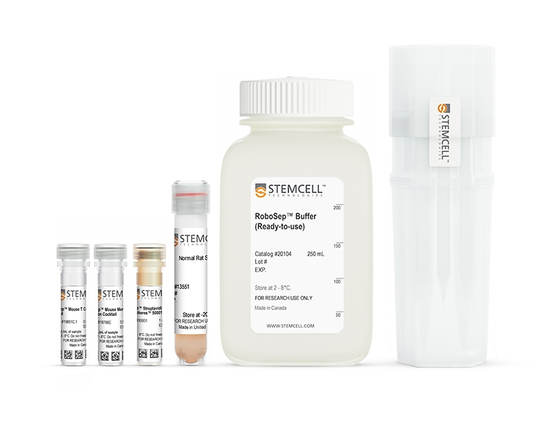
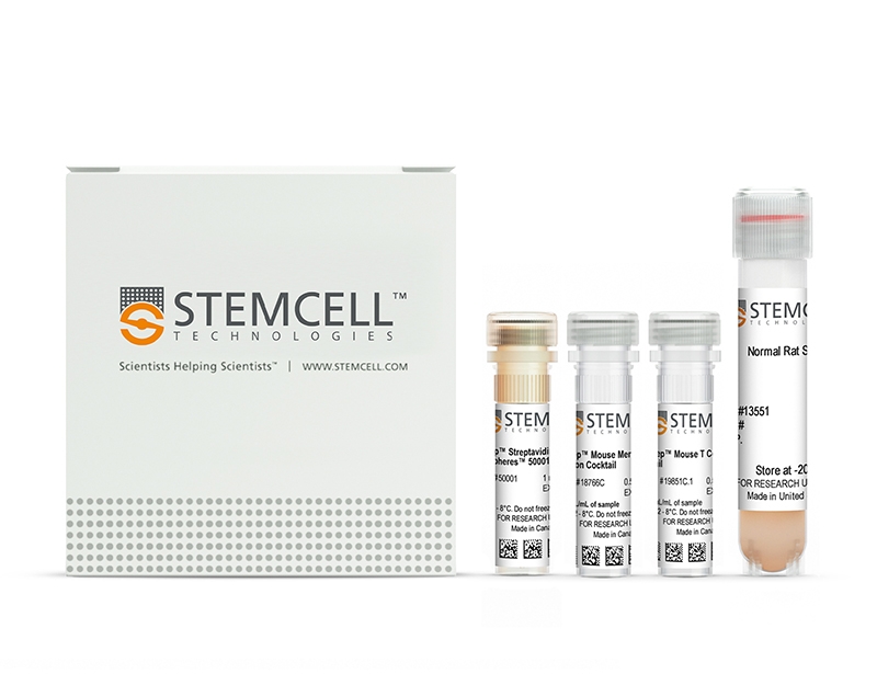
Components
- EasySep™ Mouse Pan-Naïve T Cell Isolation Kit (Catalog #19848)
- EasySep™ Mouse T Cell Isolation Cocktail, 0.5 mL
- EasySep™ Mouse Memory T Cell Depletion Cocktail, 0.5 mL
- EasySep™ Streptavidin RapidSpheres™ 50001, 1 mL
- Normal Rat Serum, 2 mL
- RoboSep™ Mouse Pan-Naïve T Cell Isolation Kit (Catalog #19848RF)
- EasySep™ Mouse T Cell Isolation Cocktail, 0.5 mL
- EasySep™ Mouse Memory T Cell Depletion Cocktail, 0.5 mL
- EasySep™ Streptavidin RapidSpheres™ 50001, 1 mL
- Normal Rat Serum, 2 mL
- RoboSep™ Buffer (Catalog #20104)
- RoboSep™ Filter Tips (Catalog #20125)
Magnet Compatibility
• EasySep™ Magnet (Catalog #18000)
• “The Big Easy” EasySep™ Magnet (Catalog #18001)
• EasyPlate™ EasySep™ Magnet (Catalog 18102)
• EasyEights™ EasySep™ Magnet (Catalog #18103)
• RoboSep™-S (Catalog #21000)
Subtype
Cell Isolation Kits
Cell Type
T Cells
Species
Mouse
Sample Source
Other, Spleen
Selection Method
Negative
Application
Cell Isolation
Brand
EasySep, RoboSep
Area of Interest
Immunology
技术资料
| Document Type | 产品名称 | Catalog # | Lot # | 语言 |
|---|---|---|---|---|
| Product Information Sheet | EasySep™ Mouse Pan-Naïve T Cell Isolation Kit | 19848 | All | English |
| Product Information Sheet | RoboSep™ Mouse Pan-Naïve T Cell Isolation Kit | 19848RF | All | English |
| Safety Data Sheet 1 | EasySep™ Mouse Pan-Naïve T Cell Isolation Kit | 19848 | All | English |
| Safety Data Sheet 2 | EasySep™ Mouse Pan-Naïve T Cell Isolation Kit | 19848 | All | English |
| Safety Data Sheet 3 | EasySep™ Mouse Pan-Naïve T Cell Isolation Kit | 19848 | All | English |
| Safety Data Sheet 1 | RoboSep™ Mouse Pan-Naïve T Cell Isolation Kit | 19848RF | All | English |
| Safety Data Sheet 2 | RoboSep™ Mouse Pan-Naïve T Cell Isolation Kit | 19848RF | All | English |
| Safety Data Sheet 3 | RoboSep™ Mouse Pan-Naïve T Cell Isolation Kit | 19848RF | All | English |
数据及文献
Data
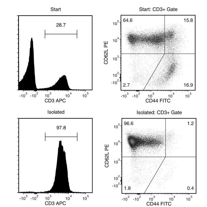
Figure 1. Typical EasySep™ Mouse Pan-Naïve T Cell Isolation Profile
Starting with mouse splenocytes from an uninfected mouse, the pan-naïve T cell (CD3+CD44-/lowCD62Lhigh) content of the isolated fraction typically ranges from 90 - 97%. In the example above, the final purities of the start and isolated fractions are 18.5% and 94.5%, respectively.
Publications (2)
Nature 2019 apr
Nuclear positioning facilitates amoeboid migration along the path of least resistance.
Abstract
Abstract
During metazoan development, immune surveillance and cancer dissemination, cells migrate in complex three-dimensional microenvironments1-3. These spaces are crowded by cells and extracellular matrix, generating mazes with differently sized gaps that are typically smaller than the diameter of the migrating cell4,5. Most mesenchymal and epithelial cells and some-but not all-cancer cells actively generate their migratory path using pericellular tissue proteolysis6. By contrast, amoeboid cells such as leukocytes use non-destructive strategies of locomotion7, raising the question how these extremely fast cells navigate through dense tissues. Here we reveal that leukocytes sample their immediate vicinity for large pore sizes, and are thereby able to choose the path of least resistance. This allows them to circumnavigate local obstacles while effectively following global directional cues such as chemotactic gradients. Pore-size discrimination is facilitated by frontward positioning of the nucleus, which enables the cells to use their bulkiest compartment as a mechanical gauge. Once the nucleus and the closely associated microtubule organizing centre pass the largest pore, cytoplasmic protrusions still lingering in smaller pores are retracted. These retractions are coordinated by dynamic microtubules; when microtubules are disrupted, migrating cells lose coherence and frequently fragment into migratory cytoplasmic pieces. As nuclear positioning in front of the microtubule organizing centre is a typical feature of amoeboid migration, our findings link the fundamental organization of cellular polarity to the strategy of locomotion.
The Journal of Immunology 2015
Skin Metabolites Define a New Paradigm in the Localization of Skin Tropic Memory T Cells
Abstract
Abstract
The localization of memory T cells to human skin is essential for long-term immune surveillance and the maintenance of barrier integrity. The expression of CCR8 during naive T cell activation is controlled by skin-specific factors derived from epidermal keratinocytes and not by resident dendritic cells. In this study, we show that the CCR8-inducing factors are heat stable and protease resistant and include the vitamin D3 metabolite 1α,25-dihydroxyvitamin D3 and PGE2. The effect of either metabolite alone on CCR8 expression was weak, whereas their combination resulted in robust CCR8 expression. Elevation of intracellular cAMP was essential because PGE2 could be substituted with the adenylyl cyclase agonist forskolin, and CCR8 expression was sensitive to protein kinase A inhibition. For effective induction, exposure of naive T cells to these epidermal factors needed to occur either prior to or during T cell activation even though CCR8 was only detected 4-5 d later in proliferating T cells. The importance of tissue environments in maintaining cellular immune surveillance networks within distinct healthy tissues provides a paradigm shift in adaptive immunity. Epidermal-derived vitamin D3 metabolites and PGs provide an essential cue for the localization of CCR8(+) immune surveillance T cells within healthy human skin.