
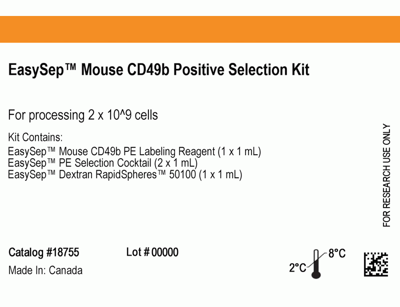
EasySep™ Mouse CD49b Positive Selection Kit
Immunomagnetic positive selection cell isolation kit
概要
The EasySep™ Mouse CD49b Positive Selection Kit is designed to isolate CD49b+ cells from single-cell suspensions of splenocytes or other tissues by positive selection. The cocktail contains a combination of PE-labeled antibodies that is directed against CD49b, which are then recognized by Tetrameric Antibody Complexes that are directed against PE and dextran. Labeled cells are bound to magnetic particles and separated using an EasySep™ magnet without the use of columns. Cells of interest remain in the tube while unwanted cells are poured off.
Advantages
• Fast and easy-to-use
• Up to 90% purity
• No columns required
• Up to 90% purity
• No columns required
Components
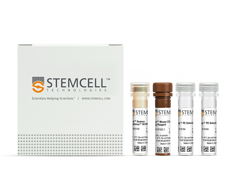
- EasySep™ Mouse CD49b Positive Selection Kit (Catalog #18755)
- EasySep™ Mouse CD49b PE Labeling Reagent, 1 mL
- EasySep™ PE Selection Cocktail, 2 x 1 mL
- EasySep™ Dextran RapidSpheres™ 50100, 1 mL
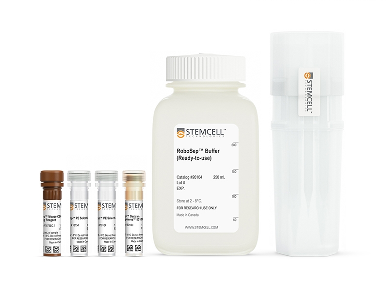
- RoboSep™ Mouse CD49b Positive Selection Kit with Filter Tips (Catalog #18755RF)
- EasySep™ Mouse CD49b PE Labeling Reagent, 1 mL
- EasySep™ PE Selection Cocktail, 2 x 1 mL
- EasySep™ Dextran RapidSpheres™ 50100, 1 mL
- RoboSep™ Buffer (Catalog #20104)
- RoboSep™ Filter Tips (Catalog #20125) x 2
Magnet Compatibility
• EasySep™ Magnet (Catalog #18000)
• “The Big Easy” EasySep™ Magnet (Catalog #18001)
• RoboSep™-S (Catalog #21000)
Subtype
Cell Isolation Kits
Cell Type
NK Cells
Species
Mouse
Sample Source
Other, Spleen
Selection Method
Positive
Application
Cell Isolation
Brand
EasySep, RoboSep
Area of Interest
Immunology
技术资料
| Document Type | 产品名称 | Catalog # | Lot # | 语言 |
|---|---|---|---|---|
| Product Information Sheet | EasySep™ Mouse CD49b Positive Selection Kit | 18755 | 17M87097 or higher | English |
| Product Information Sheet | RoboSep™ Mouse CD49b Positive Selection Kit with Filter Tips | 18755RF | 17M87097 or higher | English |
| Safety Data Sheet | EasySep™ Mouse CD49b Positive Selection Kit | 18755 | All | English |
| Safety Data Sheet | RoboSep™ Mouse CD49b Positive Selection Kit with Filter Tips | 18755RF | All | English |
数据及文献
Data
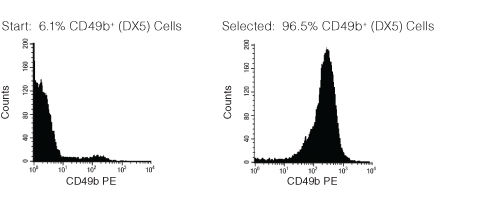
Figure 1. FACS Histogram Results with EasySep™ Mouse CD49b Positive Selection Kit
Starting with mouse splenocytes, the CD49b+ (DX5) cell content of the selected cells typically ranges from 88 - 97%.
Publications (5)
Blood 2009 SEP
Long-term protection from syngeneic acute lymphoblastic leukemia by CpG ODN-mediated stimulation of innate and adaptive immune responses.
Abstract
Abstract
Acute lymphoblastic leukemia (ALL) is the most common childhood cancer and remains a major cause of mortality in children with recurrent disease and in adults. Despite observed graft-versus-leukemia effects after stem cell transplantation, successful immune therapies for ALL have proven elusive. We previously reported immunostimulatory oligodeoxynucleotides containing CpG motifs (CpG ODN) enhance allogeneic T(h)1 responses and reduce leukemic burden of primary human ALL xenografts. To further the development of CpG ODN as a novel ALL therapy, we investigated the antileukemia activity induced by CpG ODN in a transplantable syngeneic pre-B ALL model. CpG ODN induced early killing of leukemia by innate immune effectors both in vitro and in vivo. Mice were treated with CpG ODN starting 7 days after injection with leukemia to mimic a minimal residual disease state and achieved T cell-dependent remissions of more than 6 months. In addition, mice in remission after CpG ODN treatment were protected from leukemia rechallenge, and adoptive transfer of T cells from mice in remission conferred protection against leukemia growth. To our knowledge, this is the first demonstration that CpG ODN induce a durable remission and ongoing immune-mediated protection in ALL, suggesting this treatment may have clinical utility in patients with minimal residual disease.
Journal of immunology (Baltimore, Md. : 1950) 2009 SEP
IL-33 activates unprimed murine basophils directly in vitro and induces their in vivo expansion indirectly by promoting hematopoietic growth factor production.
Abstract
Abstract
IL-33, a new member of the IL-1 family, has been described as an important inducer of Th2 cytokines and mediator of inflammatory responses. In this study, we demonstrate that murine basophils sorted directly from the bone marrow, without prior exposure to IL-3 or Fc(epsilon)R cross-linking, respond to IL-33 alone by producing substantial amounts of histamine, IL-4, and IL-6. These cells express ST2 constitutively and generate a cytokine profile that differs from their IL-3-induced counterpart by a preferential production of IL-6. In vivo, IL-33 promotes basophil expansion in the bone marrow (BM) through an indirect mechanism of action depending on signaling through the beta(c) chain shared by receptors for IL-3, GM-CSF, and IL-5. IL-3 can still signal through its specific beta(IL-3) chain in these mutant mice, which implies that it is not the unique growth-promoting mediator in this setup, but requires IL-5 and/or GMCSF. Our results support a major role of the latter growth factor, which is readily generated by total BM cells as well as sorted basophils in response to IL-33 along with low amounts of IL-3. Furthermore, GM-CSF amplifies IL-3-induced differentiation of basophils from BM cells, whereas IL-5 that is also generated in vivo, affects neither their functions nor their growth in vitro or in vivo. In conclusion, our data provide the first evidence that IL-33 not only activates unprimed basophils directly, but also promotes their expansion in vivo through induction of GM-CSF and IL-3.
Journal of immunology (Baltimore, Md. : 1950) 2007 FEB
Activating Ly-49 receptors regulate LFA-1-mediated adhesion by NK cells.
Abstract
Abstract
NK cells are important for innate resistance to tumors and viruses. Engagement of activating Ly-49 receptors expressed by NK cells leads to rapid NK cell activation resulting in target cell lysis and cytokine production. The ITAM-containing DAP12 adapter protein stably associates with activating Ly-49 receptors, and couples receptor recognition with generation of NK responses. Activating Ly-49s are potent stimulators of murine NK cell functions, yet how they mediate such activities is not well understood. We demonstrate that these receptors trigger LFA-1-dependent tight conjugation between NK cells and target cells. Furthermore, we show that activating Ly-49 receptor engagement leads to rapid DAP12-dependent up-regulation of NK cell LFA-1 adhesiveness to ICAM-1 that is also dependent on tyrosine kinases of the Syk and Src families. These results indicate for the first time that activating Ly-49s control adhesive properties of LFA-1, and by DAP12-dependent inside-out signaling. Ly-49-driven mobilization of LFA-1 adhesive function may represent a fundamental proximal event during NK cell interactions with target cells involving activating Ly-49 receptors, leading to target cell death.
Blood 2005 SEP
Contribution of Hfe expression in macrophages to the regulation of hepatic hepcidin levels and iron loading.
Abstract
Abstract
Hereditary hemochromatosis (HH), an iron overload disease associated with mutations in the HFE gene, is characterized by increased intestinal iron absorption and consequent deposition of excess iron, primarily in the liver. Patients with HH and Hfe-deficient (Hfe-/-) mice manifest inappropriate expression of the iron absorption regulator hepcidin, a peptide hormone produced by the liver in response to iron loading. In this study, we investigated the contribution of Hfe expression in macrophages to the regulation of liver hepcidin levels and iron loading. We used bone marrow transplantation to generate wild-type (wt) and Hfe-/- mice chimeric for macrophage Hfe gene expression. Reconstitution of Hfe-deficient mice with wt bone marrow resulted in augmented capacity of the spleen to store iron and in significantly decreased liver iron loading, accompanied by a significant increase of hepatic hepcidin mRNA levels. Conversely, wt mice reconstituted with Hfe-deficient bone marrow had a diminished capacity to store iron in the spleen but no significant alterations of liver iron stores or hepcidin mRNA levels. Our results suggest that macrophage Hfe participates in the regulation of splenic and liver iron concentrations and liver hepcidin expression.
Journal of immunology (Baltimore, Md. : 1950) 2004 SEP
Cutting edge: murine dendritic cells require IL-15R alpha to prime NK cells.
Abstract
Abstract
NK cells protect hosts against viral pathogens and transformed cells, and dendritic cells (DCs) play important roles in activating NK cells. We now find that murine IL-15Ralpha-deficient DCs fail to support NK cell cytolytic activity and elaboration of IFN-gamma, despite the fact that these DCs express normal levels of costimulatory molecules and IL-12. By contrast, IL-15Ralpha expression on NK cells is entirely dispensable for their activation by DCs. In addition, blockade with anti-IL-15Ralpha and anti-IL-2Rbeta but not anti-IL-2Ralpha-specific Abs prevents NK cell activation by wild-type DCs. Finally, presentation of IL-15 by purified IL-15Ralpha/Fc in trans synergizes with IL-12 to support NK cell priming. These findings suggest that murine DCs require IL-15Ralpha to present IL-15 in trans to NK cells during NK cell priming.