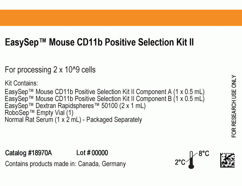
EasySep™ Mouse CD11b Positive Selection Kit II
Immunomagnetic positive selection cell isolation kit
概要
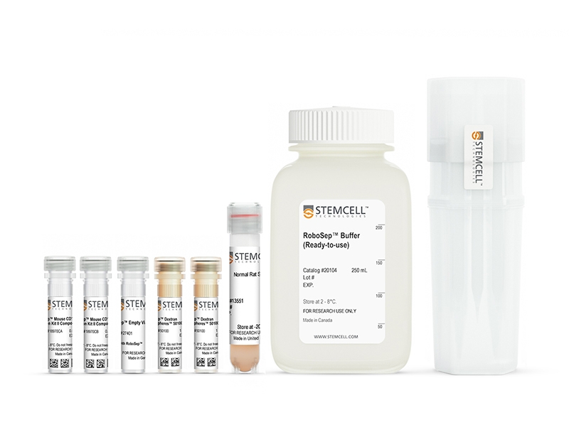
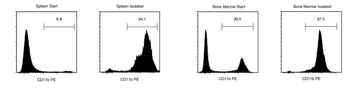
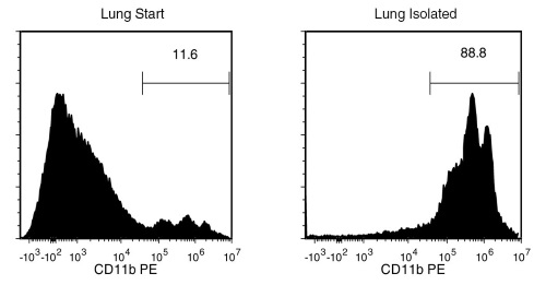
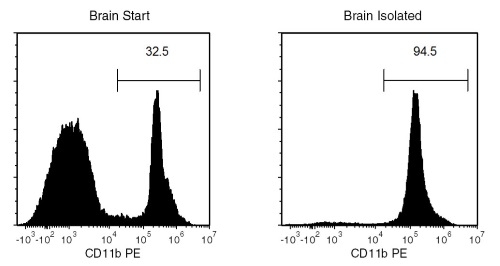
The EasySep™ Mouse CD11b Positive Selection Kit II isolates highly purified CD11b+ cells from mouse splenocytes, bone marrow, lungs, brains or other tissues by immunomagnetic positive selection. Desired cells are targeted with antibodies and magnetic particles, and isolated without columns using an EasySep™ magnet. Unwanted cells are simply poured off, while desired cells remain in the tube. Isolated cells are immediately ready for downstream applications such as flow cytometry, culture, and cell-based experiments.
This product replaces the EasySep™ Mouse CD11b Positive Selection Kit (Catalog #18770) for even faster cell isolations and does not result in the labeling of isolated cells with PE.
• Fast and easy-to-use
• Up to 95% purity (for bone marrow, purity can be up to 99%)
• No columns required
• Isolated cells are not fluorochrome-labeled
• EasySep™ Magnet (Catalog #18000)
• “The Big Easy” EasySep™ Magnet (Catalog #18001)
• EasyEights™ EasySep™ Magnet (Catalog #18103)
• RoboSep™-S (Catalog #21000)
Bone Marrow, Lung, Other, Spleen
数据及文献
Publications (4)
Scientific reports 2020 jul
Bioluminescence for in vivo detection of cell-type-specific inflammation in a mouse model of uveitis.
S. John et al.
Abstract
This study reports the use of cell-type-specific in vivo bioluminescence to measure intraocular immune cell population dynamics during the course of inflammation in a mouse model of uveitis. Transgenic lines expressing luciferase in inflammatory cell subsets (myeloid cells, T cells, and B cells) were generated and ocular bioluminescence was measured serially for 35 days following uveitis induction. Ocular leukocyte populations were identified using flow cytometry and compared to the ocular bioluminescence profile. Acute inflammation is neutrophilic (75{\%} of ocular CD45 + cells) which is reflected by a significant increase in ocular bioluminescence in one myeloid reporter line on day 2. By day 7, the ocular T cell population increases to 50{\%} of CD45 + cells, leading to a significant increase in ocular bioluminescence in the T cell reporter line. While initially negligible ({\textless} 1{\%} of CD45 + cells), the ocular B cell population increases to {\textgreater} 4{\%} by day 35. This change is reflected by a significant increase in the ocular bioluminescence of the B cell reporter line starting on day 28. Our data demonstrates that cell-type-specific in vivo bioluminescence accurately detects changes in multiple intraocular immune cell populations over time in experimental uveitis. This assay could also be useful in other inflammatory disease models.
European journal of medicinal chemistry 2020 jan
Tuning isoform selectivity and bortezomib sensitivity with a new class of alkenyl indene PDI inhibitor.
R. M. Robinson et al.
Abstract
Protein disulfide isomerase (PDI, PDIA1) is an emerging therapeutic target in oncology. PDI inhibitors have demonstrated a unique propensity to selectively induce apoptosis in cancer cells and overcome resistance to existing therapies, although drug candidates have not yet progressed to the stage of clinical development. We recently reported the discovery of lead indene compound E64FC26 as a potent pan-PDI inhibitor that enhances the cytotoxic effects of proteasome inhibitors in panels of Multiple Myeloma (MM) cells and MM mouse models. An extensive medicinal chemistry program has led to the generation of a diverse library of indene-containing molecules with varying degrees of proteasome inhibitor potentiating activity. These compounds were generated by a novel nucleophilic aromatic ring cyclization and dehydration reaction from the precursor ketones. The results provide detailed structure activity relationships (SAR) around this indene pharmacophore and show a high degree of correlation between potency of PDI inhibition and bortezomib (Btz) potentiation in MM cells. Inhibition of PDI leads to ER and oxidative stress characterized by the accumulation of misfolded poly-ubiquitinated proteins and the induction of UPR biomarkers ATF4, CHOP, and Nrf2. This work characterizes the synthesis and SAR of a new chemical class and further validates PDI as a therapeutic target in MM as a single agent and in combination with proteasome inhibitors.
Nature 2018 SEP
Clearance of senescent glial cells prevents tau-dependent pathology and cognitive decline.
T. J. Bussian et al.
Abstract
Cellular senescence, which is characterized by an irreversible cell-cycle arrest1 accompanied by a distinctive secretory phenotype2, can be induced through various intracellular and extracellular factors. Senescent cells that express the cell cycle inhibitory protein p16INK4A have been found to actively drive naturally occurring age-related tissue deterioration3,4 and contribute to several diseases associated with ageing, including atherosclerosis5 and osteoarthritis6. Various markers of senescence have been observed in patients with neurodegenerative diseases7-9; however, a role for senescent cells in the aetiology of these pathologies is unknown. Here we show a causal link between the accumulation of senescent cells and cognition-associated neuronal loss. We found that the MAPTP301SPS19 mouse model of tau-dependent neurodegenerative disease10 accumulates p16INK4A-positive senescent astrocytes and microglia. Clearance of these cells as they arise using INK-ATTAC transgenic mice prevents gliosis, hyperphosphorylation of both soluble and insoluble tau leading to neurofibrillary tangle deposition, and degeneration of cortical and hippocampal neurons, thus preserving cognitive function. Pharmacological intervention with a first-generation senolytic modulates tau aggregation. Collectively, these results show that senescent cells have a role in the initiation and progression of tau-mediated disease, and suggest that targeting senescent cells may provide a therapeutic avenue for the treatment of these pathologies.
The Journal of experimental medicine 2016 OCT
MAIT cells promote inflammatory monocyte differentiation into dendritic cells during pulmonary intracellular infection.
Meierovics AI et al.
Abstract
Mucosa-associated invariant T (MAIT) cells are a unique innate T cell subset that is necessary for rapid recruitment of activated CD4(+) T cells to the lungs after pulmonary F. tularensis LVS infection. Here, we investigated the mechanisms behind this effect. We provide evidence to show that MAIT cells promote early differentiation of CCR2-dependent monocytes into monocyte-derived DCs (Mo-DCs) in the lungs after F. tularensis LVS pulmonary infection. Adoptive transfer of Mo-DCs to MAIT cell-deficient mice (MR1(-/-) mice) rescued their defect in the recruitment of activated CD4(+) T cells to the lungs. We further demonstrate that MAIT cell-dependent GM-CSF production stimulated monocyte differentiation in vitro, and that in vivo production of GM-CSF was delayed in the lungs of MR1(-/-) mice. Finally, GM-CSF-deficient mice exhibited a defect in monocyte differentiation into Mo-DCs that was phenotypically similar to MR1(-/-) mice. Overall, our data demonstrate that MAIT cells promote early pulmonary GM-CSF production, which drives the differentiation of inflammatory monocytes into Mo-DCs. Further, this delayed differentiation of Mo-DCs in MR1(-/-) mice was responsible for the delayed recruitment of activated CD4(+) T cells to the lungs. These findings establish a novel mechanism by which MAIT cells function to promote both innate and adaptive immune responses.
View All Publications