
NeuroFluor™ NeuO

• Can be used to confirm neuronal differentiation of human pluripotent stem cell-derived neural progenitor cells
• Can be used to label neurons in live culture
• Non-toxic and non-permanent
• Simple and rapid labeling protocol
• DMSO
| Document Type | 产品名称 | Catalog # | Lot # | 语言 |
|---|---|---|---|---|
| Product Information Sheet | NeuroFluor™ NeuO | 01801 | All | English |
| Safety Data Sheet | NeuroFluor™ NeuO | 01801 | All | English |
Data
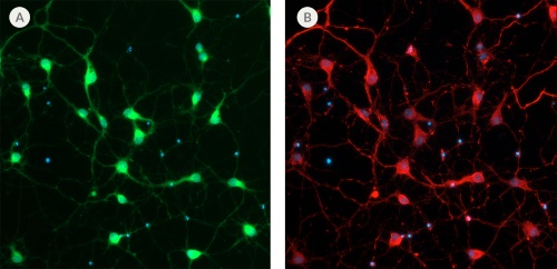
Figure 1. NeuroFluor™ NeuO Selectively Labels Primary E18 Rat Neurons
(A) Primary rat E18 cortical neurons were labeled with 0.25μM NeuroFluor™ NeuO (green) and incubated for 1 hour. Image was taken after 2 hours of incubation. (B) The same culture was later fixed and stained for β-tubulin III (red). The image shows that NeuroFluor™ NeuO specifically labels β-tubulin III-positive neurons. Nuclei are counterstained with DAPI. Images were taken at 20X magnification.
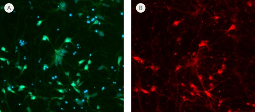
Figure 2. NeuroFluor™ NeuO Selectively Labels hPSC-Derived Neurons
(A) The neuronal precursors generated from hPSC-derived (XCL-1) neural progenitor cells were cultured in STEMdiff™ Neuron Maturation Medium. After 18 days of culture, hPSC-derived neurons were labeled with NeuroFluor™ NeuO (green). Nuclei are counterstained with DAPI. (B) The same culture was later fixed and stained with β-tubulin III (red). The image shows that NeuroFluor™ NeuO specifically labels β-tubulin III-positive neurons. Images were taken at 20X magnification.