MyoCult™-SF Expansion Supplement Kit (Human)

Serum-free supplement and attachment substrate for the derivation and expansion of human skeletal muscle progenitor cells (myoblasts)
概要
MyoCult™-SF Expansion Supplement Kit (Human) contains a serum-free supplement and attachment substrate for the culture of primary human skeletal muscle progenitor cells (myoblasts) and human pluripotent stem cell (hPSC)-derived myoblasts. The supplement must be combined with a basal medium (DMEM with 1000 mg/L D-Glucose [Catalog #36253]; sold separately) to prepare MyoCult™-SF Expansion Medium. This medium has been optimized for the derivation and expansion of human skeletal muscle progenitor cells in vitro. For primary myoblasts, cultureware must be coated with MyoCult™-SF Attachment Substrate; for hPSC-derived myoblasts, cultureware must be coated with Corning® Matrigel® hESC-Qualified Matrix. Myoblasts cultured using MyoCult™-SF Expansion Medium can be differentiated into multinucleated myotubes using MyoCult™ Differentiation Kit (Human; Catalog #05965).
Advantages
• Supports derivation and long-term expansion of human skeletal muscle progenitor cells under serum-free conditions.
• Culture-expanded skeletal muscle progenitor cells maintain differentiation potential.
• Rigorous raw material screening and quality control minimize lot-to-lot variability.
• Culture-expanded myogenic progenitor cells are compatible with MyoCult™ Differentiation Kit.
Components
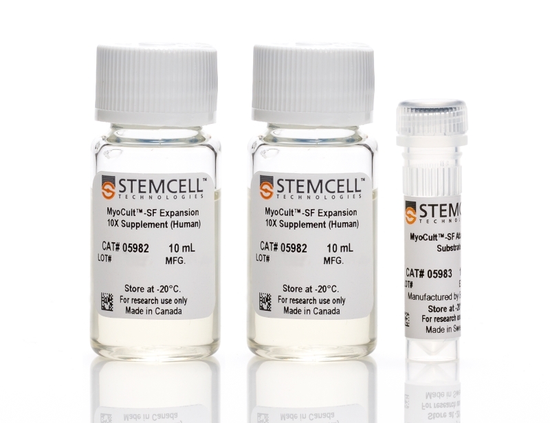
  • MyoCult™-SF Expansion 10X Supplement (Human), 2 x 10 mL
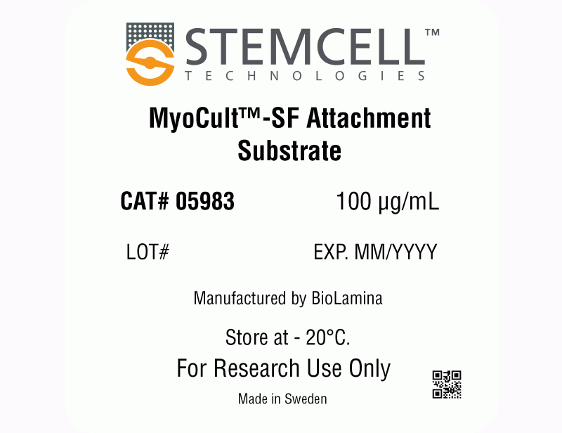
  • MyoCult™-SF Attachment Substrate, 100 µg
Subtype
Specialized Media
Cell Type
Myogenic Stem and Progenitor Cells
Species
Human
Application
Cell Culture, Expansion
Brand
MyoCult
Area of Interest
Stem Cell Biology
Formulation
Serum-Free
数据及文献

Data

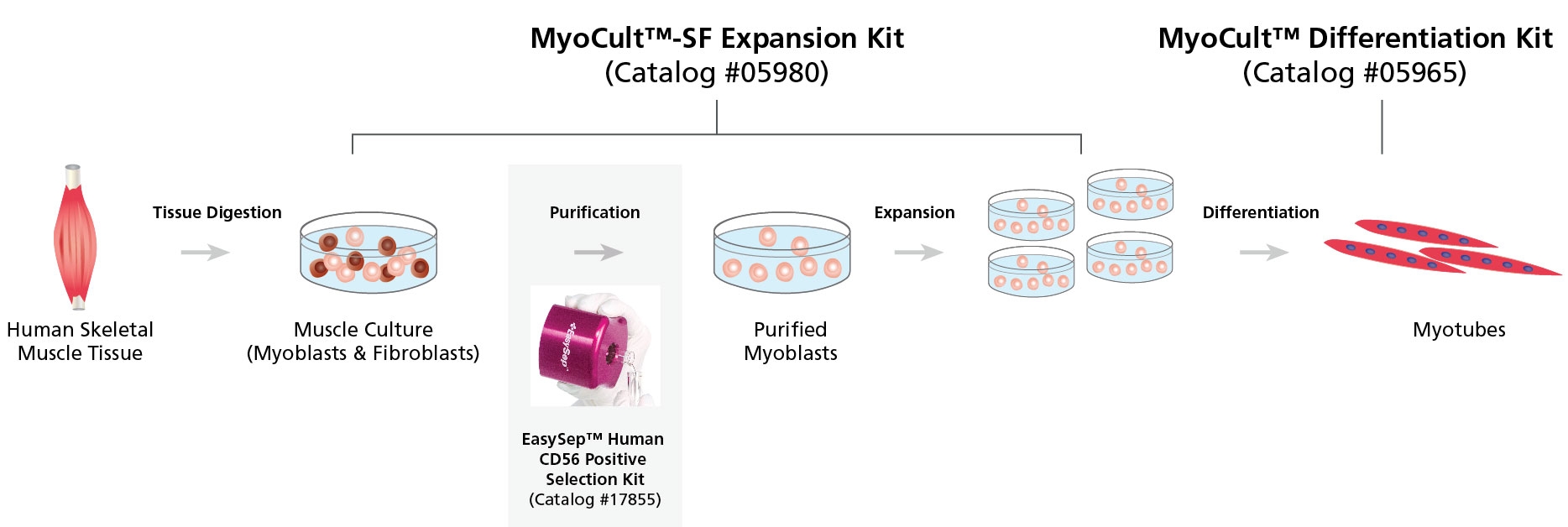
Figure 1. Workflow for the Derivation, Expansion and Differentiation of Human Myogenic Progenitor Cells

Human skeletal muscle tissue is digested into a single cell suspension and plated into MyoCult™-SF Expansion Medium. Myoblasts are then enriched by cell sorting or using a selection kit such as the EasySep™ Human CD56 Positive Selection Kit (Catalog #17855). Purified myoblasts are then culture-expanded or differentiated into myotubes for further studies. Myoblasts can be derived and expanded, using the MyoCult™-SF Expansion Kit (Catalog #05980) and then differentiated using the MyoCult™ Differentiation Kit (Catalog #05965).
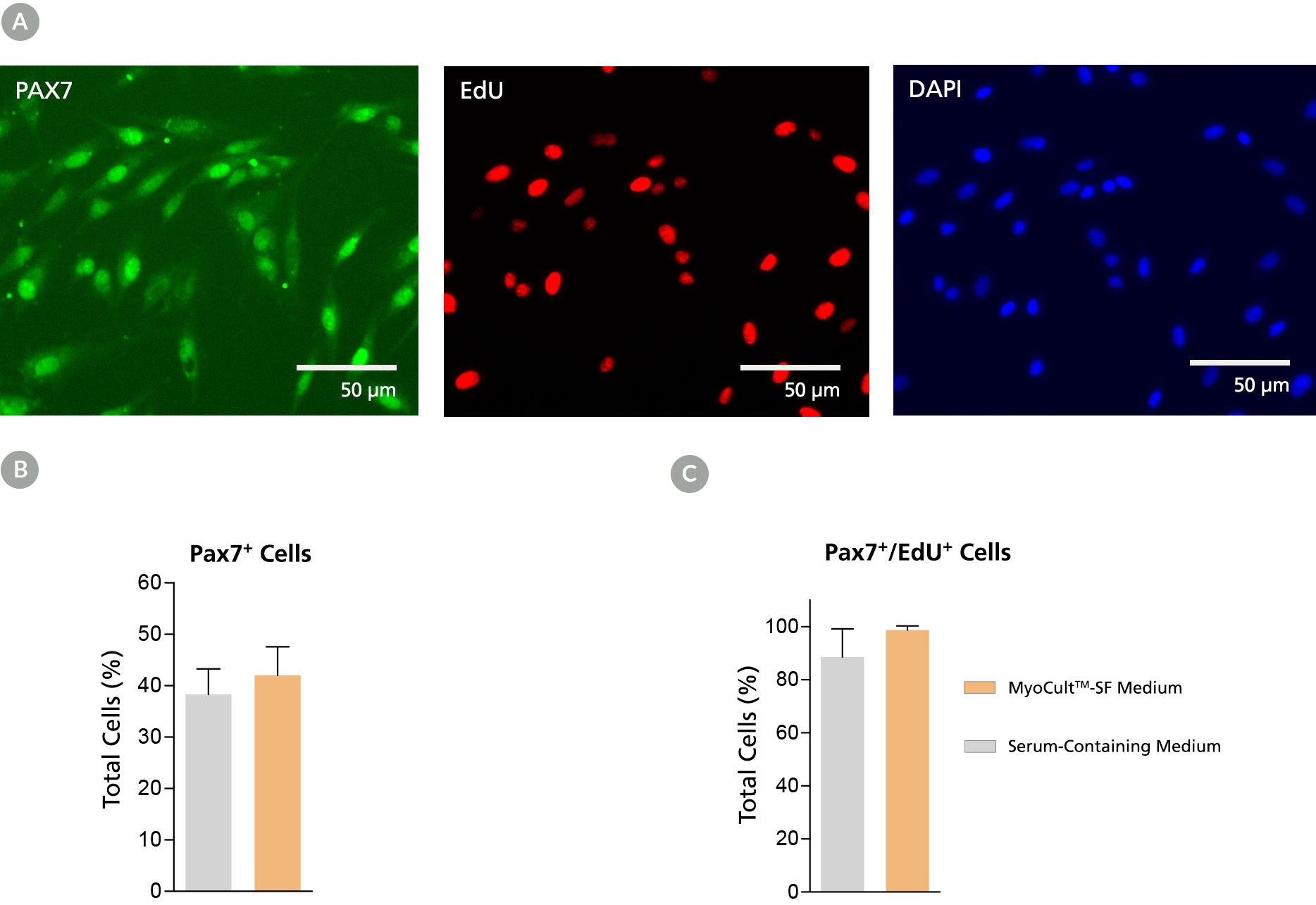
Figure 2. Derivation of PAX7+ Myogenic Progenitor Cells in MyoCult™-SF Expansion Medium from Human Skeletal Muscle Tissue

Myogenic progenitor cells were derived from human skeletal muscle tissue and culture-expanded using the MyoCult™-SF Expansion Kit or a serum-containing medium. (A) Following 6 days of expansion, myoblasts were fixed and immunostained for PAX7 (green), a thymine analogue (EdU; red) and nuclei (DAPI; blue). Total percentage of cells expressing (B) PAX7 or (C) PAX7 and EdU were quantified (n = 4). Data was generated and used with permission by Dr. Penny M. Gilbert’s Lab, Department of Biochemistry, University of Toronto.
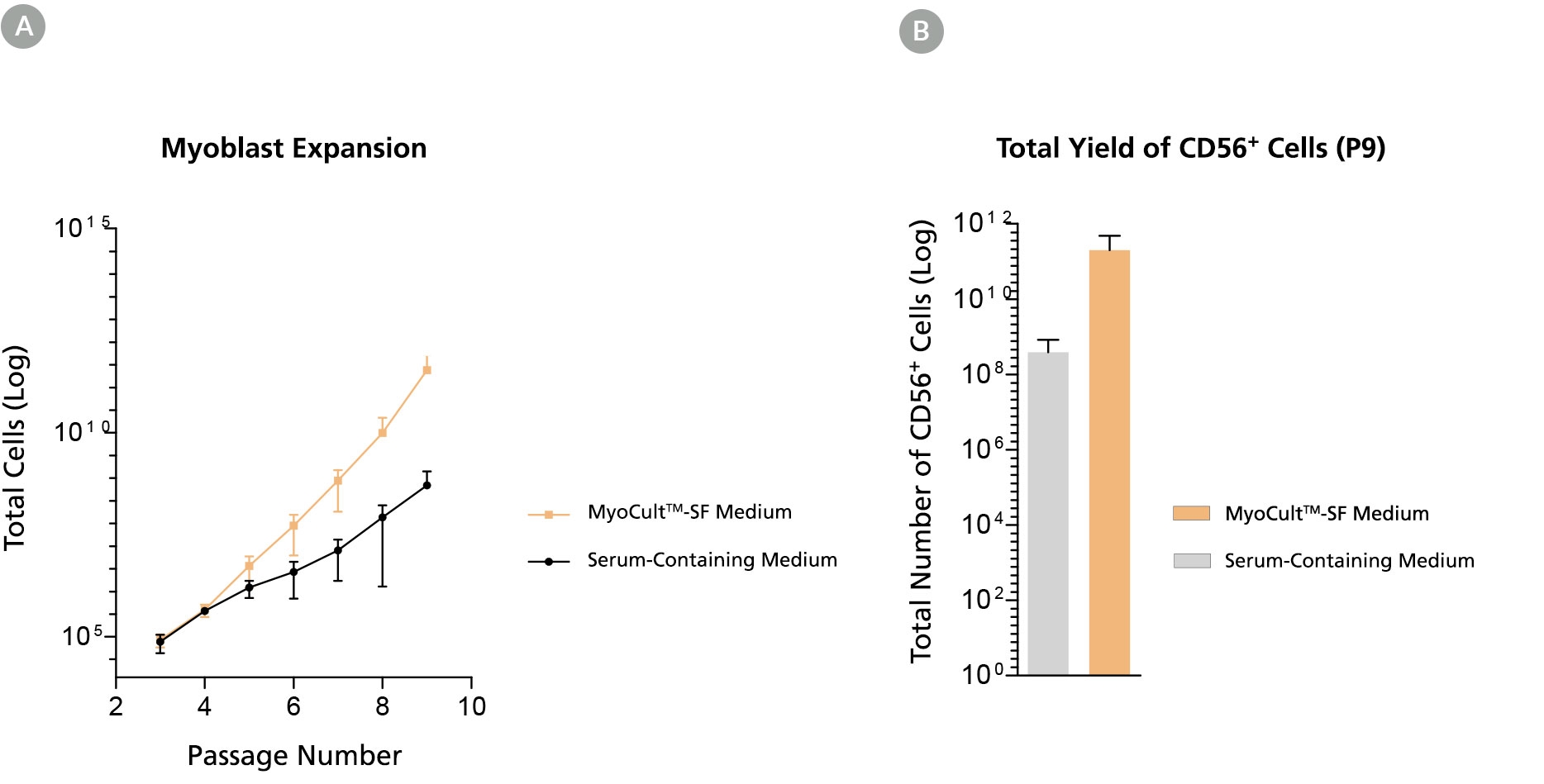
Figure 3. Long-Term Expansion of Myoblasts were Observed When Using MyoCult™-SF Expansion Kit

Myoblasts were culture-expanded in MyoCult™-SF Expansion Medium or in serum-containing medium. (A) Myoblasts culture-expanded in MyoCult™-SF Expansion Medium displayed superior expansion rate than when cells were expanded in serum-containing medium (n=3). (B) Expansion of myoblasts in MyoCult™-SF Expansion Medium generates a greater yield of CD56+ cells after 9 passages compared to myoblasts cultured in serum-containing medium (P9; n = 2). Data was generated and used with permission by Dr. Penny M. Gilbert’s Lab, Department of Biochemistry, University of Toronto.
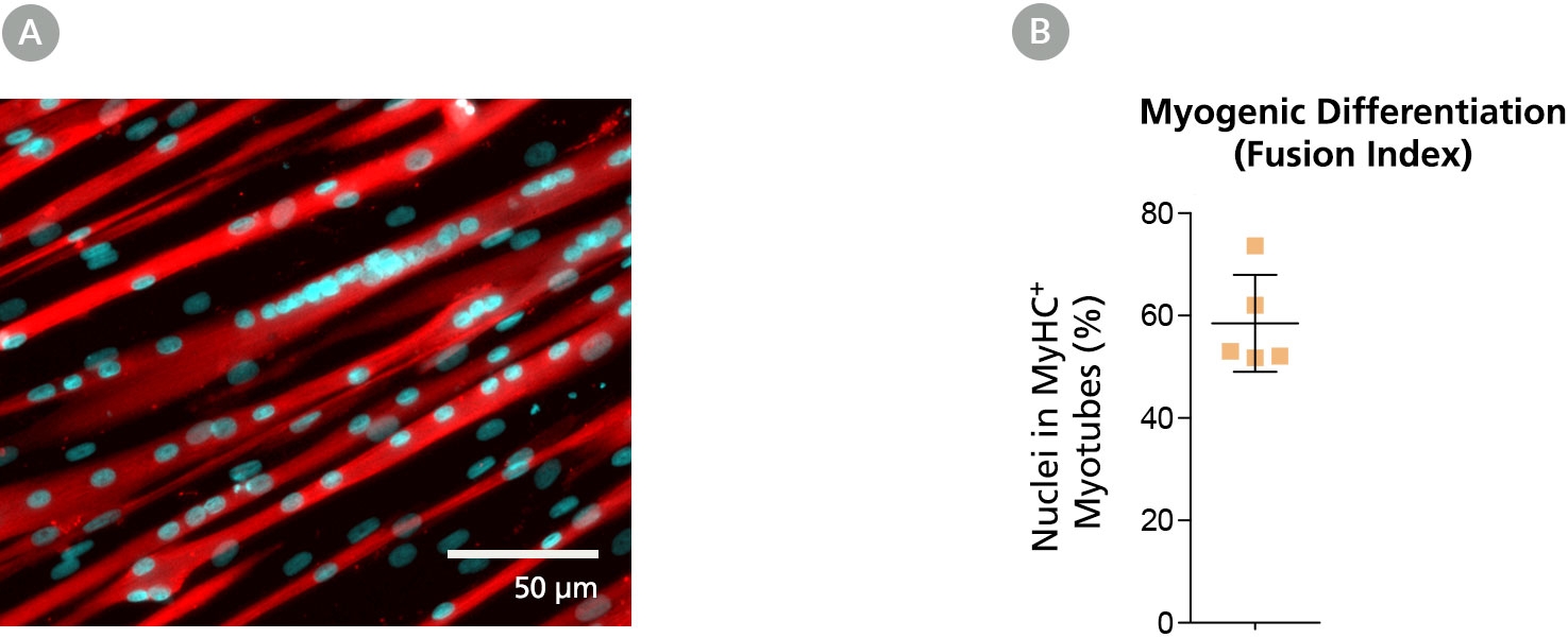
Figure 4. Myoblasts Culture-Expanded in MyoCult™-SF Expansion Medium Maintain Differentiation and Transplantation Potential in Mice

Myoblasts derived and expanded in MyoCult™-SF Expansion Medium were further differentiated into myotubes using the MyoCult™ Differentiation Kit (Catalog #05965) at passage 5. (A) Myotubes differentiated from myoblasts were immunostained for myosin heavy chain (MyHC; red) and nuclei (DAPI; blue). (B) Fusion index displaying approximately 60% of total nuclei were found within myotubes expressing MyHC. Each donor is indicated by a yellow square. Data from 5 independent donors.
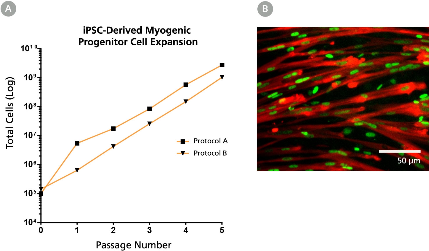
Figure 5. The MyoCult™-SF Expansion Medium Supports Expansion of iPSC-Derived Myogenic Progenitor Cells

(A) iPSC-derived myogenic progenitor cells displayed long-term expansion when using the MyoCult™ Expansion Kit. iPSC-derived myogenic progenitor cells were generated using Protocol A (Chal et al. from Nature Protocols (2016)) and Protocol B (Xi et al. Cell Rep (2017)). (B) Culture-expanded iPSC-derived myogenic progenitor cells expressed MyHC (red) and MyoD (green).

Publications (3)

ACS chemical neuroscience 2020 Differentiation of Intrafusal Fibers from Human Induced Pluripotent Stem Cells. A. Col\'on et al.

Abstract

Human-based body-on-a-chip" technology provides powerful platforms in developing models for drug evaluation and disease evaluations in phenotypic models. Induced pluripotent stem cells (iPSCs) are ideal cell sources for generating different cell types for these in vitro functional systems and recapitulation of the neuromuscular reflex arc would allow for the study of patient specific neuromuscular diseases. Regarding relevant afferent (intrafusal fibers sensory neurons) and efferent (extrafusal fibers motoneurons) cells in vitro differentiation of intrafusal fiber from human iPSCs has not been established. This work demonstrates a protocol for inducing an enrichment of intrafusal bag fibers from iPSCs using morphological analysis and immunocytochemistry. Phosphorylation of the ErbB2 receptors and S46 staining indicated a 3-fold increase of total intrafusal fibers further confirming the efficiency of the protocol. Integration of induced intrafusal fibers would enable more accurate reflex arc models and application of this protocol on patient iPSCs would allow for patient-specific disease modeling."
Physiological reports 2019 sep CD4+ T cell activation and associated susceptibility to HIV-1 infection in vitro increased following acute resistance exercise in human subjects. A. K. Holbrook et al.

Abstract

Early studies in exercise immunology suggested acute bouts of exercise had an immunosuppressive effect in human subjects. However, recent data, show acute bouts of combined aerobic and resistance training increase both lymphocyte activation and proliferation. We quantified resistance exercise-induced changes in the activation state of CD4+ T lymphocytes via surface protein expression and using a medically relevant model of infection (HIV-1). Using a randomized cross-over design, 10 untrained subjects completed a control and exercise session. The control session consisted of 30-min seated rest while the exercise session entailed 3 sets × 10 repetitions of back squat, leg press, and leg extensions at 70{\%} 1-RM with 2-min rest between each set. Venous blood samples were obtained pre/post each session. CD4+ T lymphocytes were isolated from whole blood by negative selection. Expression of activation markers (CD69 {\&} CD25) in both nonstimulated and stimulated (costimulation through CD3+ CD28) cells were assessed by flow cytometry. Resistance exercised-induced effects on intracellular activation was further evaluated via in vitro infection with HIV-1. Nonstimulated CD4+ T lymphocytes obtained postexercise exhibited elevated CD25 expression following 24 h in culture. Enhanced HIV-1 replication was observed in cells obtained postexercise. Our results demonstrate that an acute bout of resistance exercise increases the activation state of CD4+ T lymphocytes and results in a greater susceptibility to HIV-1 infection in vitro. These findings offer further evidence that exercise induces activation of T lymphocytes and provides a foundation for the use of medically relevant pathogens as indirect measures of intracellular activation.
Aging cell 2019 dec Metformin blunts muscle hypertrophy in response to progressive resistance exercise training in older adults: A randomized, double-blind, placebo-controlled, multicenter trial: The MASTERS trial. R. G. Walton et al.

Abstract

Progressive resistance exercise training (PRT) is the most effective known intervention for combating aging skeletal muscle atrophy. However, the hypertrophic response to PRT is variable, and this may be due to muscle inflammation susceptibility. Metformin reduces inflammation, so we hypothesized that metformin would augment the muscle response to PRT in healthy women and men aged 65 and older. In a randomized, double-blind trial, participants received 1,700 mg/day metformin (N = 46) or placebo (N = 48) throughout the study, and all subjects performed 14 weeks of supervised PRT. Although responses to PRT varied, placebo gained more lean body mass (p = .003) and thigh muscle mass (p {\textless} .001) than metformin. CT scan showed that increases in thigh muscle area (p = .005) and density (p = .020) were greater in placebo versus metformin. There was a trend for blunted strength gains in metformin that did not reach statistical significance. Analyses of vastus lateralis muscle biopsies showed that metformin did not affect fiber hypertrophy, or increases in satellite cell or macrophage abundance with PRT. However, placebo had decreased type I fiber percentage while metformin did not (p = .007). Metformin led to an increase in AMPK signaling, and a trend for blunted increases in mTORC1 signaling in response to PRT. These results underscore the benefits of PRT in older adults, but metformin negatively impacts the hypertrophic response to resistance training in healthy older individuals. ClinicalTrials.gov Identifier: NCT02308228.
View All Publications
Top