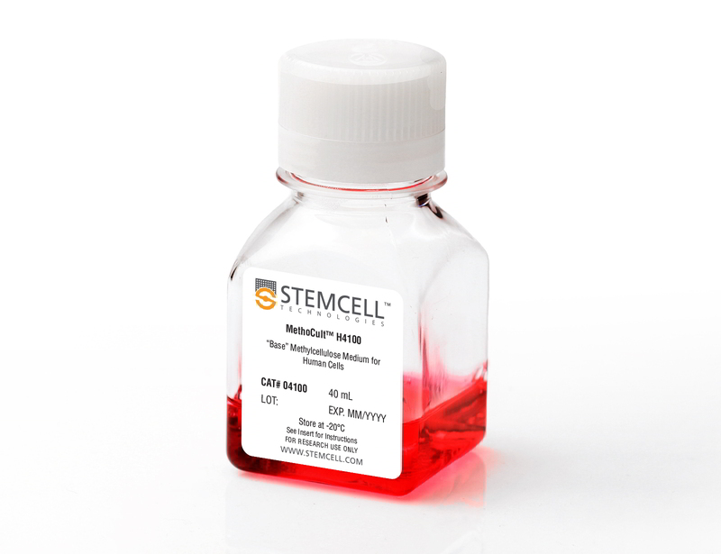
MethoCult™ H4100
Base methylcellulose medium for human cells
概要
MethoCult™ H4100 is an incomplete medium that contains 2.6% methylcellulose in Iscove's MDM. MethoCult™ H4100 is suitable for the growth and enumeration of hematopoietic progenitor cells in colony-forming unit (CFU) assays of human bone marrow, mobilized peripheral blood, peripheral blood, and cord blood samples, when the appropriate growth factors and supplements are added. This formulation does not contain serum or cytokines and can also be used as a base for cloning cell lines in semi-solid media.
Browse our Frequently Asked Questions (FAQs) on performing the CFU assay and explore its utility as part of the cell therapy workflow.
Browse our Frequently Asked Questions (FAQs) on performing the CFU assay and explore its utility as part of the cell therapy workflow.
Contains
• 2.6% Methylcellulose
• Iscove's MDM
• Iscove's MDM
Subtype
Semi-Solid Media, Specialized Media
Cell Type
Hematopoietic Stem and Progenitor Cells
Species
Human
Application
Cell Culture, Colony Assay, Functional Assay
Brand
MethoCult
Area of Interest
Stem Cell Biology
Formulation
Serum-Free
技术资料
| Document Type | 产品名称 | Catalog # | Lot # | 语言 |
|---|---|---|---|---|
| Product Information Sheet | MethoCult™ H4100 | 04100 | All | English |
| Manual | MethoCult™ H4100 | 04100 | All | English |
| Safety Data Sheet | MethoCult™ H4100 | 04100 | All | English |
数据及文献
Publications (17)
Blood 2010 JAN
High-mobility group protein HMGB2 regulates human erythroid differentiation through trans-activation of GFI1B transcription.
Abstract
Abstract
Gfi-1B is a transcriptional repressor that is crucial for erythroid differentiation: inactivation of the GFI1B gene in mice leads to embryonic death due to failure to produce differentiated red cells. Accordingly, GFI1B expression is tightly regulated during erythropoiesis, but the mechanisms involved in such regulation remain partially understood. We here identify HMGB2, a high-mobility group HMG protein, as a key regulator of GFI1B transcription. HMGB2 binds to the GFI1B promoter in vivo and up-regulates its trans-activation most likely by enhancing the binding of Oct-1 and, to a lesser extent, of GATA-1 and NF-Y to the GFI1B promoter. HMGB2 expression increases during erythroid differentiation concomitantly to the increase of GfI1B transcription. Importantly, knockdown of HMGB2 in immature hematopoietic progenitor cells leads to decreased Gfi-1B expression and impairs their erythroid differentiation. We propose that HMGB2 potentiates GATA-1-dependent transcription of GFI1B by Oct-1 and thereby controls erythroid differentiation.
Cancer research 2010 DEC
Persistent activation of the Fyn/ERK kinase signaling axis mediates imatinib resistance in chronic myelogenous leukemia cells through upregulation of intracellular SPARC.
Abstract
Abstract
SPARC is an extracellular matrix protein that exerts pleiotropic effects on extracellular matrix organization, growth factor availability, cell adhesion, differentiation, and immunity in cancer. Chronic myelogenous leukemia (CML) cells resistant to the BCR-ABL inhibitor imatinib (IM-R cells) were found to overexpress SPARC mRNA. In this study, we show that imatinib triggers SPARC accumulation in a variety of tyrosine kinase inhibitor (TKI)-resistant CML cell lines. SPARC silencing in IM-R cells restored imatinib sensitivity, whereas enforced SPARC expression in imatinib-sensitive cells promoted viability as well as protection against imatinib-mediated apoptosis. Notably, we found that the protective effect of SPARC required intracellular retention inside cells. Accordingly, SPARC was not secreted into the culture medium of IM-R cells. Increased SPARC expression was intimately linked to persistent activation of the Fyn/ERK kinase signaling axis. Pharmacologic inhibition of this pathway or siRNA-mediated knockdown of Fyn kinase resensitized IM-R cells to imatinib. In support of our findings, increased levels of SPARC mRNA were documented in blood cells from CML patients after 1 year of imatinib therapy compared with initial diagnosis. Taken together, our results highlight an important role for the Fyn/ERK signaling pathway in imatinib-resistant cells that is driven by accumulation of intracellular SPARC.
Blood 2009 FEB
Small molecule XIAP inhibitors cooperate with TRAIL to induce apoptosis in childhood acute leukemia cells and overcome Bcl-2-mediated resistance.
Abstract
Abstract
Defects in apoptosis contribute to poor outcome in pediatric acute lymphoblastic leukemia (ALL), calling for novel strategies that counter apoptosis resistance. Here, we demonstrate for the first time that small molecule inhibitors of the antiapoptotic protein XIAP cooperate with TRAIL to induce apoptosis in childhood acute leukemia cells. XIAP inhibitors at subtoxic concentrations, but not a structurally related control compound, synergize with TRAIL to trigger apoptosis and to inhibit clonogenic survival of acute leukemia cells, whereas they do not affect viability of normal peripheral blood lymphocytes, suggesting some tumor selectivity. Analysis of signaling pathways reveals that XIAP inhibitors enhance TRAIL-induced activation of caspases, loss of mitochondrial membrane potential, and cytochrome c release in a caspase-dependent manner, indicating that they promote a caspase-dependent feedback mitochondrial amplification loop. Of note, XIAP inhibitors even overcome Bcl-2-mediated resistance to TRAIL by enhancing Bcl-2 cleavage and Bak conformational change. Importantly, XIAP inhibitors kill leukemic blasts from children with ALL ex vivo and cooperate with TRAIL to induce apoptosis. In vivo, they significantly reduce leukemic burden in a mouse model of pediatric ALL engrafted in non-obese diabetic/severe combined immunodeficient (NOD/SCID) mice. Thus, XIAP inhibitors present a promising novel approach for apoptosis-based therapy of childhood ALL.
Molecular and cellular biology 2008 DEC
Anchorage-independent growth of pocket protein-deficient murine fibroblasts requires bypass of G2 arrest and can be accomplished by expression of TBX2.
Abstract
Abstract
Mouse embryonic fibroblasts (MEFs) deficient for pocket proteins (i.e., pRB/p107-, pRB/p130-, or pRB/p107/p130-deficient MEFs) have lost proper G(1) control and are refractory to Ras(V12)-induced senescence. However, pocket protein-deficient MEFs expressing Ras(V12) were unable to exhibit anchorage-independent growth or to form tumors in nude mice. We show that depending on the level of pocket proteins, loss of adhesion induces G(1) and G(2) arrest, which could be alleviated by overexpression of the TBX2 oncogene. TBX2-induced transformation occurred only in the absence of pocket proteins and could be attributed to downregulation of the p53/p21(CIP1) pathway. Our results show that a balance between the pocket protein and p53 pathways determines the level of transformation of MEFs by regulating cyclin-dependent kinase activities. Since transformation of human fibroblasts also requires ablation of both pathways, our results imply that the mechanisms underlying transformation of human and mouse cells are not as different as previously claimed.
The Journal of experimental medicine 2007 SEP
Unregulated actin polymerization by WASp causes defects of mitosis and cytokinesis in X-linked neutropenia.
Abstract
Abstract
Specific mutations in the human gene encoding the Wiskott-Aldrich syndrome protein (WASp) that compromise normal auto-inhibition of WASp result in unregulated activation of the actin-related protein 2/3 complex and increased actin polymerizing activity. These activating mutations are associated with an X-linked form of neutropenia with an intrinsic failure of myelopoiesis and an increase in the incidence of cytogenetic abnormalities. To study the underlying mechanisms, active mutant WASp(I294T) was expressed by gene transfer. This caused enhanced and delocalized actin polymerization throughout the cell, decreased proliferation, and increased apoptosis. Cells became binucleated, suggesting a failure of cytokinesis, and micronuclei were formed, indicative of genomic instability. Live cell imaging demonstrated a delay in mitosis from prometaphase to anaphase and confirmed that multinucleation was a result of aborted cytokinesis. During mitosis, filamentous actin was abnormally localized around the spindle and chromosomes throughout their alignment and separation, and it accumulated within the cleavage furrow around the spindle midzone. These findings reveal a novel mechanism for inhibition of myelopoiesis through defective mitosis and cytokinesis due to hyperactivation and mislocalization of actin polymerization.
Blood 2006 SEP
Human CD34+ cells expressing the inv(16) fusion protein exhibit a myelomonocytic phenotype with greatly enhanced proliferative ability.
Abstract
Abstract
The t(16:16) and inv(16) are associated with FAB M4Eo myeloid leukemias and result in fusion of the CBFB gene to the MYH11 gene (encoding smooth muscle myosin heavy chain [SMMHC]). Knockout of CBFbeta causes embryonic lethality due to lack of definitive hematopoiesis. Although knock-in of CBFB-MYH11 is not sufficient to cause disease, expression increases the incidence of leukemia when combined with cooperating events. Although mouse models are valuable tools in the study of leukemogenesis, little is known about the contribution of CBFbeta-SMMHC to human hematopoietic stem and progenitor cell self-renewal. We introduced the CBFbeta-MYH11 cDNA into human CD34+ cells via retroviral transduction. Transduced cells displayed an initial repression of progenitor activity but eventually dominated the culture, resulting in the proliferation of clonal populations for up to 7 months. Long-term cultures displayed a myelomonocytic morphology while retaining multilineage progenitor activity and engraftment in NOD/SCID-B2M-/- mice. Progenitor cells from long-term cultures showed altered expression of genes defining inv(16) identified in microarray studies of human patient samples. This system will be useful in examining the effects of CBFbeta-SMMHC on gene expression in the human preleukemic cell, in characterizing the effect of this oncogene on human stem cell biology, and in defining its contribution to the development of leukemia.


