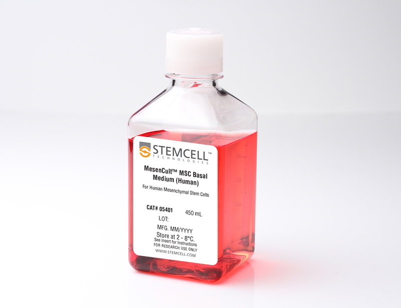
MesenCult™ MSC Basal Medium (Human)
Basal medium for human mesenchymal stem cells
概要
MesenCult™ MSC Basal Medium (Human) is a standardized basal medium designed to be supplemented with MesenCult™ Mesenchymal Stem Cell Stimulatory Supplement (Human; Catalog #05402) for the in vitro culture of human mesenchymal stem cells (MSCs). MesenCult™ MSC Basal Medium is a component of MesenCult™ Proliferation Kit (Human; Catalog #05411), and is also available separately.
Subtype
Basal Media
Cell Type
Mesenchymal Stem and Progenitor Cells
Species
Human
Application
Cell Culture, Colony Assay, Expansion
Brand
MesenCult
Area of Interest
Stem Cell Biology
技术资料
| Document Type | 产品名称 | Catalog # | Lot # | 语言 |
|---|---|---|---|---|
| Product Information Sheet 1 | MesenCult™ MSC Basal Medium (Human) | 05401 | Lot 19D102073A or higher | English |
| Product Information Sheet 2 | MesenCult™ MSC Basal Medium (Human) | 05401 | 19A99097 or lower | English |
| Safety Data Sheet | MesenCult™ MSC Basal Medium (Human) | 05401 | All | English |
数据及文献
Publications (48)
Cells 2020 mar
FAK Deficiency in Bone Marrow Stromal Cells Alters Their Homeostasis and Drives Abnormal Proliferation and Differentiation of Haematopoietic Stem Cells.
Abstract
Abstract
Emerging evidence indicates that in myelodysplastic syndromes (MDS), the bone marrow (BM) microenvironment may also contribute to the ineffective, malignant haematopoiesis in addition to the intrinsic abnormalities of haematopoietic stem precursor cells (HSPCs). The BM microenvironment influences malignant haematopoiesis through indirect mechanisms, but the processes by which the BM microenvironment directly contributes to MDS initiation and progression have not yet been elucidated. Our previous data showed that BM-derived stromal cells (BMSCs) from MDS patients have an abnormal expression of focal adhesion kinase (FAK). In this study, we characterise the morpho-phenotypic features and the functional alterations of BMSCs from MDS patients and in FAK knock-downed HS-5 cells. The decreased expression of FAK or its phosphorylated form in BMSCs from low-risk (LR) MDS directly correlates with BMSCs' functional deficiency and is associated with a reduced level of haemoglobin. The downregulation of FAK in HS-5 cells alters their morphology, proliferation, and differentiation capabilities and impairs the expression of several adhesion molecules. In addition, we examine the CD34+ healthy donor (HD)-derived HSPCs' properties when co-cultured with FAK-deficient BMSCs. Both abnormal proliferation and the impaired erythroid differentiation capacity of HD-HSPCs were observed. Together, these results demonstrate that stromal adhesion mechanisms mediated by FAK are crucial for regulating HSPCs' homeostasis.
Life sciences 2020 jul
Icariin protects rabbit BMSCs against OGD-induced apoptosis by inhibiting ERs-mediated autophagy via MAPK signaling pathway.
Abstract
Abstract
Stem cell therapy is widely employed in treating osteoarthritis (OA), and bone marrow-derived mesenchymal stem cells (BMSCs) has gradually become the most attractive new method for treating OA due to the benefit for cartilage tissue repair. However, the apoptosis in the neural stem cell transplantation severely decreases repairing efficacy. Icariin has been reported to exert multiple effects on BMSCs, including its proliferation, osteogenic, and chondrogenic differentiation. However, its effects on the injury induced by oxygen, glucose and serum deprivation (OGD) remains unknown. We prospectively investigated the role of ICA on rabbit BMSCs under conditions of OGD. Firstly, BMSCs were cultured under conditions of OGD, ICA relieved OGD-induced cell damage by promoting cell proliferation and suppressing apoptosis. Secondly, Markers of endoplasmic reticulum stress (ERs), ER stress IRE-1 pathway, and autophagy were both inhibited by ICA via inhibition of phosphor-extracellular regulated protein kinases (p-ERKs), p-P38, p-c-Jun N-terminal kinase (p-JNK) or si-MAPK. Finally, decrease of ERs marker levels enhanced protective effect of ICA against OGD-induced injury by limiting apoptosis and autophagy. Moreover, an autophagy inhibitor (3-methyladenine: 3-MA) contributed to a synergistic effect in conjunction with ICA, in promoting cell proliferation, suggesting that ICA exerts anti-ERs and anti-autophagy effects in OGD-treated BMSCs. Therefore, ICA protected rabbit BMSCs from OGD-induced apoptosis through inhibitory regulation of ERs-mediated autophagy related to the MAPK signaling pathway, which provided insights for a potential therapeutic strategy in OA.
PloS one 2019
Isolation of adipose tissue derived regenerative cells from human subcutaneous tissue with or without the use of an enzymatic reagent.
Abstract
Abstract
Freshly isolated, uncultured, autologous adipose derived regenerative cells (ADRCs) have emerged as a promising tool for regenerative cell therapy. The Transpose RT system (InGeneron, Inc., Houston, TX, USA) is a system for isolating ADRCs from adipose tissue, commercially available in Europe as a CE-marked medical device and under clinical evaluation in the United States. This system makes use of the proprietary, enzymatic Matrase Reagent for isolating cells. The present study addressed the question whether the use of Matrase Reagent influences cell yield, cell viability, live cell yield, biological characteristics, physiological functions or structural properties of the ADRCs in final cell suspension. Identical samples of subcutaneous adipose tissue from 12 subjects undergoing elective lipoplasty were processed either with or without the use of Matrase Reagent. Then, characteristics of the ADRCs in the respective final cell suspensions were evaluated. Compared to non-enzymatic isolation, enzymatic isolation resulted in approximately twelve times higher mean cell yield (i.e., numbers of viable cells/ml lipoaspirate) and approximately 16 times more colony forming units. Despite these differences, cells isolated from lipoaspirate both with and without the use of Matrase Reagent were independently able to differentiate into cells of all three germ layers. This indicates that biological characteristics, physiological functions or structural properties relevant for the intended use were not altered or induced using Matrase Reagent. A comprehensive literature review demonstrated that isolation of ADRCs from lipoaspirate using the Transpose RT system and the Matrase Reagent results in the highest viable cell yield among published data regarding isolation of ADRCs from lipoaspirate.
Radiotherapy and oncology : journal of the European Society for Therapeutic Radiology and Oncology 2019
1-(4-nitrobenzenesulfonyl)-4-penylpiperazine increases the number of Peyer's patch-associated regenerating crypts in the small intestines after radiation injury.
Abstract
Abstract
OBJECTIVE Exposure to lethal doses of radiation has severe effects on normal tissues. Exposed individuals experience a plethora of symptoms in different organ systems including the gastrointestinal (GI) tract, summarized as Acute Radiation Syndrome (ARS). There are currently no approved drugs for mitigating GI-ARS. A recent high-throughput screen performed at the UCLA Center for Medical Countermeasures against Radiation identified compounds containing sulfonylpiperazine groups with radiation mitigation properties to the hematopoietic system and the gut. Among these 1-[(4-Nitrophenyl)sulfonyl]-4-phenylpiperazine (Compound {\#}5) efficiently mitigated gastrointestinal ARS. However, the mechanism of action and target cells of this drug is still unknown. In this study we examined if Compound {\#}5 affects gut-associated lymphoid tissue (GALT) with its subepithelial domes called Peyer's patches. METHODS C3H mice were irradiated with 0 or 12 Gy total body irradiation (TBI). A single dose of Compound {\#}5 or solvent was administered subcutaneously 24 h later. 48 h after irradiation the mice were sacrificed, and the guts examined for changes in the number of visible Peyer's patches. In some experiments the mice received 4 daily injections of treatment and were sacrificed 96 h after TBI. For immune histochemistry gut tissues were fixed in formalin and embedded in paraffin blocks. Sections were stained with H{\&}E, anti-Ki67 or a TUNEL assay to assess the number of regenerating crypts, mitotic and apoptotic indices. Cells isolated from Peyer's patches were subjected to immune profiling using flow cytometry. RESULTS Compound {\#}5 significantly increased the number of visible Peyer's patches when compared to its control in non-irradiated and irradiated mice. Additionally, assessment of total cells per Peyer's patch isolated from these mice demonstrated an overall increase in the total number of Peyer's patch cells per mouse in Compound {\#}5-treated mice. In non-irradiated animals the number of CD11bhigh in Peyer's patches increased significantly. These Compound {\#}5-driven increases did not coincide with a decrease in apoptosis or an increase in proliferation in the germinal centers inside Peyer's patches 24 h after drug treatment. A single dose of Compound {\#}5 significantly increased the number of CD45+ cells after 12 Gy TBI. Importantly, 96 h after 12 Gy TBI Compound {\#}5 induced a significant rise in the number of visible Peyer's patches and the number of Peyer's patch-associated regenerating crypts. CONCLUSION In summary, our study provides evidence that Compound {\#}5 leads to an influx of immune cells into GALT, thereby supporting crypt regeneration preferentially in the proximity of Peyer's patches.
Molecular cancer therapeutics 2019
A Unique Nonsaccharide Mimetic of Heparin Hexasaccharide Inhibits Colon Cancer Stem Cells via p38 MAP Kinase Activation.
Abstract
Abstract
Targeting of cancer stem cells (CSC) is expected to be a paradigm-shifting approach for the treatment of cancers. Cell surface proteoglycans bearing sulfated glycosaminoglycan (GAG) chains are known to play a critical role in the regulation of stem cell fate. Here, we show for the first time that G2.2, a sulfated nonsaccharide GAG mimetic (NSGM) of heparin hexasaccharide, selectively inhibits colonic CSCs in vivo G2.2-reduced CSCs (CD133+/CXCR4+, Dual hi) induced HT-29 and HCT 116 colon xenografts' growth in a dose-dependent fashion. G2.2 also significantly delayed the growth of colon xenograft further enriched in CSCs following oxaliplatin and 5-fluorouracil treatment compared with vehicle-treated xenograft controls. In fact, G2.2 robustly inhibited CSCs' abundance (measured by levels of CSC markers, e.g., CD133, DCMLK1, LGR5, and LRIG1) and self-renewal (quaternary spheroids) in colon cancer xenografts. Intriguingly, G2.2 selectively induced apoptosis in the Dual hi CSCs in vivo eluding to its CSC targeting effects. More importantly, G2.2 displayed none to minimal toxicity as observed through morphologic and biochemical studies of vital organ functions, blood coagulation profile, and ex vivo analyses of normal intestinal (and bone marrow) progenitor cell growth. Through extensive in vitro, in vivo, and ex vivo mechanistic studies, we showed that G2.2's inhibition of CSC self-renewal was mediated through activation of p38$\alpha$, uncovering important signaling that can be targeted to deplete CSCs selectively while minimizing host toxicity. Hence, G2.2 represents a first-in-class (NSGM) anticancer agent to reduce colorectal CSCs.
Cellular reprogramming 2014 FEB
Mesenchymal Derivatives of Genetically Unstable Human Embryonic Stem Cells Are Maintained Unstable but Undergo Senescence in Culture As Do Bone Marrow–Derived Mesenchymal Stem Cells
Abstract
Abstract
Recurrent chromosomal alterations have been repeatedly reported in cultured human embryonic stem cells (hESCs). The effects of these alterations on the capability of pluripotent cells to differentiate and on growth potential of their specific differentiated derivatives remain unclear. Here, we report that the hESC lines HUES-7 and -9 carrying multiple chromosomal alterations produce in vitro mesenchymal stem cells (MSCs) that show progressive growth arrest and enter senescence after 15 and 16 passages, respectively. There was no difference in their proliferative potential when compared with bone marrow-derived MSCs. Array comparative genomic hybridization analysis (aCGH) of hESCs and their mesenchymal derivatives revealed no significant differences in chromosomal alterations, suggesting that genetically altered hESCs are not selected out during differentiation. Our findings indicate that genetically unstable hESCs maintain their capacity to differentiate in vitro into MSCs, which exhibit an in vitro growth pattern of normal MSCs and not that of transformed cells.


