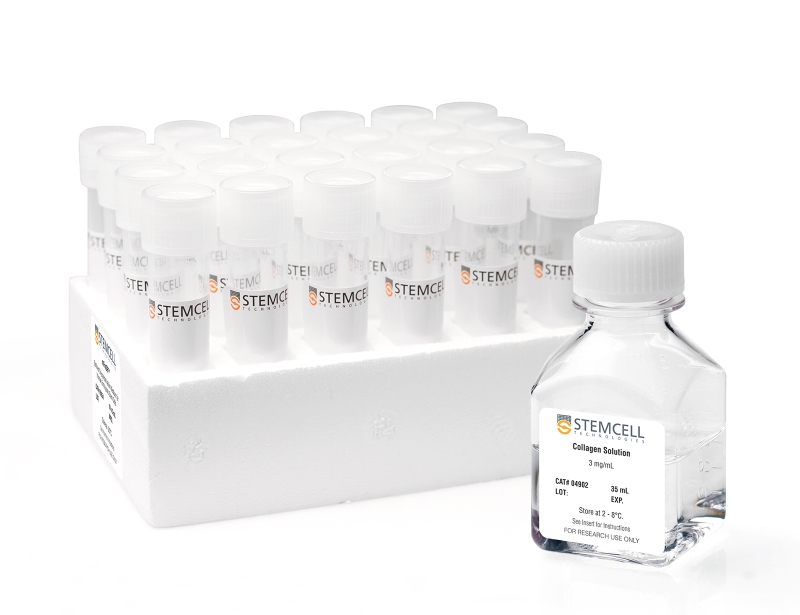
MegaCult™-C Collagen and Medium with Cytokines
Collagen and medium with cytokines for human CFU-Mk assays
概要
MegaCult™-C Collagen and Medium with Cytokines kit contains medium and collagen solution for colony-forming unit (CFU) assays of human megakaryocyte progenitor cells (CFU-Mk). The medium contains recombinant human (rh) IL-3, IL-6, and thrombopoietin.
MegaCult™-C medium is optimized for the growth of CFU-Mk in human bone marrow, mobilized peripheral blood, and cord blood samples. It is suitable for use with CD34+ enriched cells, mononuclear cells, and cells isolated by other purification methods.
MegaCult™-C medium is optimized for the growth of CFU-Mk in human bone marrow, mobilized peripheral blood, and cord blood samples. It is suitable for use with CD34+ enriched cells, mononuclear cells, and cells isolated by other purification methods.
Components
- MegaCult™-C Collagen and Medium with Cytokines (Catalog #04961)
- Collagen Solution, 35 mL (Catalog #04902)
- MegaCult™-C Medium with Cytokines, 24 x 2 mL (Catalog #04901)
Subtype
Semi-Solid Media, Specialized Media
Cell Type
Hematopoietic Stem and Progenitor Cells
Species
Human
Application
Cell Culture, Colony Assay, Functional Assay
Brand
MegaCult
Area of Interest
Stem Cell Biology
Formulation
Serum-Free
技术资料
| Document Type | 产品名称 | Catalog # | Lot # | 语言 |
|---|---|---|---|---|
| Product Information Sheet 1 | MegaCult™-C Collagen and Medium with Cytokines | 04961 | All | English |
| Product Information Sheet 2 | MegaCult™-C Collagen and Medium with Cytokines | 04961 | All | English |
| Product Information Sheet | MegaCult™-C Medium with Cytokines | 04901 | All | English |
| Product Information Sheet | Collagen Solution | 04902 | All | English |
| Manual | MegaCult™-C Collagen and Medium with Cytokines | 04961 | All | English |
| Safety Data Sheet | MegaCult™-C Collagen and Medium with Cytokines | 04961 | All | English |
| Safety Data Sheet | MegaCult™-C Medium with Cytokines | 04901 | All | English |
| Safety Data Sheet | Collagen Solution | 04902 | All | English |
数据及文献
Publications (23)
Blood 2009 APR
Mef2C is a lineage-restricted target of Scl/Tal1 and regulates megakaryopoiesis and B-cell homeostasis.
Abstract
Abstract
The basic helix-loop-helix transcription factor stem cell leukemia gene (Scl) is a master regulator for hematopoiesis essential for hematopoietic specification and proper differentiation of the erythroid and megakaryocyte lineages. However, the critical downstream targets of Scl remain undefined. Here, we identified a novel Scl target gene, transcription factor myocyte enhancer factor 2 C (Mef2C) from Scl(fl/fl) fetal liver progenitor cell lines. Analysis of Mef2C(-/-) embryos showed that Mef2C, in contrast to Scl, is not essential for specification into primitive or definitive hematopoietic lineages. However, adult VavCre(+)Mef2C(fl/fl) mice exhibited platelet defects similar to those observed in Scl-deficient mice. The platelet counts were reduced, whereas platelet size was increased and the platelet shape and granularity were altered. Furthermore, megakaryopoiesis was severely impaired in vitro. Chromatin immunoprecipitation microarray hybridization analysis revealed that Mef2C is directly regulated by Scl in megakaryocytic cells, but not in erythroid cells. In addition, an Scl-independent requirement for Mef2C in B-lymphoid homeostasis was observed in Mef2C-deficient mice, characterized as severe age-dependent reduction of specific B-cell progenitor populations reminiscent of premature aging. In summary, this work identifies Mef2C as an integral member of hematopoietic transcription factors with distinct upstream regulatory mechanisms and functional requirements in megakaryocyte and B-lymphoid lineages.
Blood 2008 MAY
Transgenic expression of JAK2V617F causes myeloproliferative disorders in mice.
Abstract
Abstract
The JAK2(V617F) mutation was found in most patients with myeloproliferative disorders (MPDs), including polycythemia vera, essential thrombocythemia, and primary myelofibrosis. We have generated transgenic mice expressing the mutated enzyme in the hematopoietic system driven by a vav gene promoter. The mice are viable and fertile. One line of the transgenic mice, which expressed a lower level of JAK2(V617F), showed moderate elevations of blood cell counts, whereas another line with a higher level of JAK2(V617F) expression displayed marked increases in blood counts and developed phenotypes that closely resembled human essential thrombocythemia and polycythemia vera. The latter line of mice also developed primary myelofibrosis-like symptoms as they aged. The transgenic mice showed erythroid, megakaryocytic, and granulocytic hyperplasia in the bone marrow and spleen, displayed splenomegaly, and had reduced levels of plasma erythropoietin and thrombopoietin. They possessed an increased number of hematopoietic progenitor cells in peripheral blood, spleen, and bone marrow, and these cells formed autonomous colonies in the absence of growth factors and cytokines. The data show that JAK2(V617F) can cause MPDs in mice. Our study thus provides a mouse model to study the pathologic role of JAK2(V617F) and to develop treatment for MPDs.
Journal of immunological methods 2008 MAR
Increased production of megakaryocytes near purity from cord blood CD34+ cells using a short two-phase culture system.
Abstract
Abstract
Expansion of hematopoietic progenitor cells (HPC) ex vivo remains an important focus in fundamental and clinical research. The aim of this study was to determine whether the implementation of such expansion phase in a two-phase culture strategy prior to the induction of megakaryocyte (Mk) differentiation would increase the yield of Mks produced in cultures. Toward this end, we first characterized the functional properties of five cytokine cocktails to be tested in the expansion phase on the growth and differentiation kinetics of CD34+-enriched cells, and on their capacity to expand clonogenic progenitors in cultures. Three of these cocktails were chosen based on their reported ability to induce HPC expansion ex vivo, while the other two represented new cytokine combinations. These analyses revealed that none of the cocktails tested could prevent the differentiation of CD34+ cells and the rapid expansion of lineage-positive cells. Hence, we sought to determine the optimum length of time for the expansion phase that would lead to the best final Mk yields. Despite greater expansion of CD34+ cells and overall cell growth with a longer expansion phase, the optimal length for the expansion phase that provided greater Mk yield at near maximal purity was found to be 5 days. Under such settings, two functionally divergent cocktails were found to significantly increase the final yield of Mks. Surprisingly, these cocktails were either deprived of thrombopoietin or of stem cell factor, two cytokines known to favor megakaryopoiesis and HPC expansion, respectively. Based on these results, a short resource-efficient two-phase culture protocol for the production of Mks near purity (textgreater95%) from human CD34+ CB cells has been established.
The Journal of clinical investigation 2007 SEP
Tescalcin is an essential factor in megakaryocytic differentiation associated with Ets family gene expression.
Abstract
Abstract
We show here that the process of megakaryocytic differentiation requires the presence of the recently discovered protein tescalcin. Tescalcin is dramatically upregulated during the differentiation and maturation of primary megakaryocytes or upon PMA-induced differentiation of K562 cells. This upregulation requires sustained signaling through the ERK pathway. Overexpression of tescalcin in K562 cells initiates events of spontaneous megakaryocytic differentiation, such as expression of specific cell surface antigens, inhibition of cell proliferation, and polyploidization. Conversely, knockdown of this protein in primary CD34+ hematopoietic progenitors and cell lines by RNA interference suppresses megakaryocytic differentiation. In cells lacking tescalcin, the expression of Fli-1, Ets-1, and Ets-2 transcription factors, but not GATA-1 or MafB, is blocked. Thus, tescalcin is essential for the coupling of ERK cascade activation with the expression of Ets family genes in megakaryocytic differentiation.
Blood 2007 JUL
A novel fusion of RBM6 to CSF1R in acute megakaryoblastic leukemia.
Abstract
Abstract
Activated tyrosine kinases have been frequently implicated in the pathogenesis of cancer, including acute myeloid leukemia (AML), and are validated targets for therapeutic intervention with small-molecule kinase inhibitors. To identify novel activated tyrosine kinases in AML, we used a discovery platform consisting of immunoaffinity profiling coupled to mass spectrometry that identifies large numbers of tyrosine-phosphorylated proteins, including active kinases. This method revealed the presence of an activated colony-stimulating factor 1 receptor (CSF1R) kinase in the acute megakaryoblastic leukemia (AMKL) cell line MKPL-1. Further studies using siRNA and a small-molecule inhibitor showed that CSF1R is essential for the growth and survival of MKPL-1 cells. DNA sequence analysis of cDNA generated by 5'RACE from CSF1R coding sequences identified a novel fusion of the RNA binding motif 6 (RBM6) gene to CSF1R gene generated presumably by a t(3;5)(p21;q33) translocation. Expression of the RBM6-CSF1R fusion protein conferred interleukin-3 (IL-3)-independent growth in BaF3 cells, and induces a myeloid proliferative disease (MPD) with features of megakaryoblastic leukemia in a murine transplant model. These findings identify a novel potential therapeutic target in leukemogenesis, and demonstrate the utility of phosphoproteomic strategies for discovery of tyrosine kinase alleles.
Stem cells (Dayton, Ohio) 2007 APR
CD41+/CD45+ cells without acetylcholinesterase activity are immature and a major megakaryocytic population in murine bone marrow.
Abstract
Abstract
Murine megakaryocytes (MKs) are defined by CD41/CD61 expression and acetylcholinesterase (AChE) activity; however, their stages of differentiation in bone marrow (BM) have not been fully elucidated. In murine lineage-negative (Lin(-))/CD45(+) BM cells, we found CD41(+) MKs without AChE activity (AChE(-)) except for CD41(++) MKs with AChE activity (AChE(+)), in which CD61 expression was similar to their CD41 level. Lin(-)/CD41(+)/CD45(+)/AChE(-) MKs could differentiate into AChE(+), with an accompanying increase in CD41/CD61 during in vitro culture. Both proplatelet formation (PPF) and platelet (PLT) production for Lin(-)/CD41(+)/CD45(+)/AChE(-) MKs were observed later than for Lin(-)/CD41(++)/CD45(+)/AChE(+) MKs, whereas MK progenitors were scarcely detected in both subpopulations. GeneChip and semiquantitative polymerase chain reaction analyses revealed that the Lin(-)/CD41(+)/CD45(+)/AChE(-) MKs are assigned at the stage between the progenitor and PPF preparation phases in respect to the many MK/PLT-specific gene expressions, including beta1-tubulin. In normal mice, the number of Lin(-)/CD41(+)/CD45(+)/AChE(-) MKs was 100 times higher than that of AChE(+) MKs in BM. When MK destruction and consequent thrombocytopenia were caused by an antitumor agent, mitomycin-C, Lin(-)/CD41(+)/CD45(+)/AChE(-) MKs led to an increase in AChE(+) MKs and subsequent PLT recovery with interleukin-11 administration. It was concluded that MKs in murine BM at least in part consist of immature Lin(-)/CD41(+)/CD45(+)/AChE(-) MKs and more differentiated Lin(-)/CD41(++)/CD45(+)/AChE(+) MKs. Immature Lin(-)/CD41(+)/CD45(+)/AChE(-) MKs are a major MK population compared with AChE(+) MKs in BM and play an important role in rapid PLT recovery in vivo.


