ImmunoCult™ Human Treg Differentiation Supplement
This product is recommended for:
• Research into the regulation of human naïve T cell differentiation and Treg function
• Development of procedures to expand and polarize naïve T cells into Tregs
• Optimized for use with ImmunoCult™-XF T Cell Expansion Medium
• Supports higher levels of memory T cells in comparison to competitor Treg differentiation kit
• Supports up to 20% higher proportions of CD4+CD25+FOXP3+T cells in comparison to competitor Treg differentiation kit
• Supplied as a 50X concentrate; after thawing and mixing, the tube contents can be added directly to medium
• All-trans retinoic acid
| Document Type | 产品名称 | Catalog # | Lot # | 语言 |
|---|---|---|---|---|
| Product Information Sheet | ImmunoCult™ Human Treg Differentiation Supplement | 10977 | All | English |
| Safety Data Sheet | ImmunoCult™ Human Treg Differentiation Supplement | 10977 | All | English |
Data
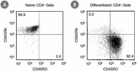
Figure 1. ImmunoCult™ Human Treg Differentiation Supplement Produces Differentiated CD4+CD45RA-CD45RO+ Cells Under Treg Polarizing Conditions
Naïve CD4+ T cells were isolated from human peripheral blood samples using the EasySep™ Human Naïve CD4+ T Cell Isolation Kit (Catalog #19555). Cells were stimulated with ImmunoCult™ Human CD3/CD28 T Cell Activator (Catalog #10971) and ImmunoCult™ Human Treg Differentiation Supplement (Catalog #10977), cultured in ImmunoCult™-XF T Cell Expansion Medium (Catalog #10981) for 7 days. Shown are flow cytometry results of cell samples stained and analyzed for expression of CD45RA and CD45RO (A) before and (B) after 7 days of culture. After 7 days the majority of Treg cells in culture display a memory T cell phenotype (CD45RA-CD45RO+).
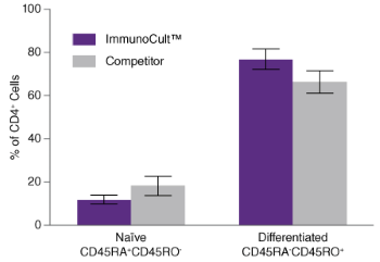
Figure 2. ImmunoCult™ Human Treg Differentiation Supplement Produces Similar Numbers of Differentiated CD4+CD45RA-CD45RO+ Cells Under Treg Polarizing Conditions Than Competitor
Naïve CD4+ T cells were isolated from human peripheral blood samples using the EasySep™ Human Naïve CD4+ T Cell Isolation Kit (Catalog #19555) and cultured with ImmunoCult™ Human CD3/CD28 T Cell Activator (Catalog #10971) and ImmunoCult™ Human Treg Differentiation Supplement (Catalog #10977) in ImmunoCult™-XF T Cell Expansion Medium (Catalog #10981) (purple), or competitor medium and activator (grey) for 5 days (based on the length of the competitor protocol). The percentage of naïve (CD45RA+CD45RO-) and differentiated (CD45RA-CD45RO+) cells after culture was compared after staining cells for CD45RA and CD45RO and analysis by flow cytometry. The percentage of cells expressing a naïve phenotype and the percentage of cells expressing a differentiated phenotype were not significantly higher with ImmunoCult™ than with competitor medium and activator. Shown are the differences in the percentage of naïve and differentiated T cells in either ImmunoCult™ (purple) and competitor (grey) culture conditions (mean ± S.E.M.; paired t-test on logit transformed data; n = 4).
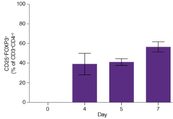
Figure 3. ImmunoCult™ Human Treg Differentiation Supplement Produces FOXP3+ Cells Under Treg Polarizing Conditions
Naïve CD4+ T cells were cultured for 7 days in ImmunoCult™ activator, supplement and medium using the protocol described in Figure 2. Cells were stained for CD4, CD4, CD25 and FOXP3 and analyzed on days 0, 4, 5 and 7 by flow cytometry. Each bar represents the mean percentage of CD3+CD4+ cells expressing both CD25 and FOXP3 (CD3+CD4+CD25+FOXP3+) (mean ± S.E.M; n = 4 – 20 donors).
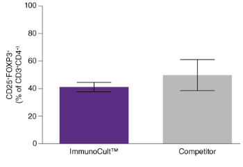
Figure 4. Culture with ImmunoCult™ Human Treg Differentiation Supplement Produces Similar Percentages of CD3+CD4+CD25+FOXP3+ Cells as Culture Under Competitor Conditions
Naïve CD4+ T cells were cultured for 5 days in ImmunoCult™ activator, supplement and medium, or competitor medium and activator using the protocol described in Figure 2. The percentage of CD25+FOXP3+ after 5 days of culture was compared after staining cells for CD3, CD4, CD25 and FOXP3 and analysis by flow cytometry. Similar percentages of CD3+CD4+ cells generated with ImmunoCult™ expressed CD25 and FOXP3 as compared to those generated under competitor conditions. Shown are the differences in the percentage of CD3+CD4+ cells expressing both CD25 and FOXP3 (CD3+CD4+CD25+FOXP3+) in either ImmunoCult™ (purple) and competitor (grey) culture conditions (mean ± S.E.M.; paired t-test on logit transformed data; n = 4 - 20).

