ImmunoCult™-XF T Cell Expansion Medium

Serum-free and xeno-free medium for the expansion of human T cells
概要
ImmunoCult™-XF T Cell Expansion Medium is a serum-free and xeno-free medium optimized for the in vitro culture and expansion of human T cells isolated from peripheral blood. Recombinant cytokines, required for the optimal growth and expansion of T cells, have not been added to ImmunoCult™-XF T Cell Expansion Medium. This allows users the flexibility to prepare medium that meets their requirements.

This product is designed for cell therapy research applications following the recommendations of USP<1043> on Ancillary Materials, and we can currently work with you to qualify this reagent under an approved investigational new drug (IND) or clinical trial application (CTA).
Advantages
• No need to supplement the medium with serum
• Supports robust T cell expansion with high viability after 10 - 12 days of culture
• Expanded T cells are able to produce cytokines including IFN-gamma and IL-4 upon restimulation
• Use with ImmunoCult™ Human T Cell Activators (Catalog #10970 and 10971) for bead-free activation of T cells
Subtype
Specialized Media
Cell Type
T Cells, T Cells, CD4+, T Cells, CD8+
Species
Human
Application
Cell Culture, Expansion
Brand
ImmunoCult
Area of Interest
Cell Therapy, Immunology
Formulation
Serum-Free, Xeno-Free
数据及文献

Data

ImmunoCult™-XF T Cell Expansion Medium Supports Faster T Cell Expansion Than Other Serum-Free and Serum-Supplemented Media

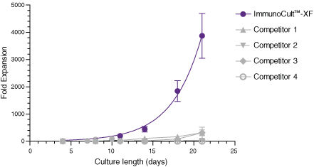
Figure 1. ImmunoCult™-XF T Cell Expansion Medium Supports Faster T Cell Expansion Than Other Serum-Free and Serum-Supplemented Media

T cells were isolated from human peripheral blood samples using the EasySep™ Human T Cell Isolation Kit (Catalog #17951), stimulated with ImmunoCult™ Human CD3/CD28/CD2 T Cell Activator (Catalog #10970), and cultured in ImmunoCult™-XF T Cell Expansion Medium supplemented with rhIL-2. T cells were stimulated with ImmunoCult™ Human CD3/CD28/CD2 T Cell Activator on day 0 and every 7 to 8 days for the duration of the culture. T cells were analyzed on days 4, 7, 8, 10, 11, 14, 18, and 21 for fold expansion relative to the initial cell seeding density. Compared to all competitor media tested, ImmunoCult™-XF T Cell Expansion Medium showed significantly higher expansion of total T cells. Competitors 1 to 4 include, in no particular order, X-VIVO™ 15 (Lonza), AIM V® Medium (Life Tech), CellGro® DC Medium (CellGenix), and RPMI 1640 + serum. Each data point represents the mean fold expansion ± S.E.M. at the specified time points (p<0.05 for ImmunoCult™-XF versus all media for days 8, 11, 14, 18, and 21, tested using two-tailed, paired t-test with unequal variance, n = 6 to 19 donors). The average fold expansion of T cells in ImmunoCult™-XF T Cell Expansion Medium were 15-fold on day 7, 80-fold on day 10, 450-fold on day 14, and 4,000-fold on day 21.

ImmunoCult™-XF T Cell Expansion Medium Supports Greater T Cell Expansion Than Other Serum-Free and Serum-Supplemented Media

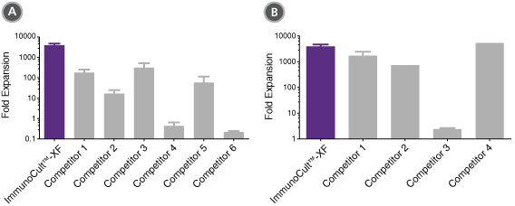
Figure 2. ImmunoCult™-XF T Cell Expansion Medium Supports Greater T Cell Expansion Than Other Serum-Free and Serum-Supplemented Media

T cells were isolated from human peripheral blood samples using the EasySep™ Human T Cell Isolation Kit (Catalog #17951), stimulated with ImmunoCult™ Human CD3/CD28/CD2 T Cell Activator (Catalog #10970), and cultured in (A) ImmunoCult™-XF T Cell Expansion Medium or serum-free competitor media with rhIL-2 in three replicate cultures per donor, or cultured in (B) ImmunoCult™-XF T Cell Expansion Medium or serum-supplemented competitor media with rhIL-2 in three replicate cultures per donor. T cells were stimulated with ImmunoCult™ Human CD3/CD28/CD2 T Cell Activator on day 0 and every 7 to 8 days for the duration of the culture. T cells were analyzed on day 21 for fold expansion relative to the initial cell seeding density.
(A) Compared to all serum-free competitor media tested, ImmunoCult™-XF T Cell Expansion Medium showed significantly higher expansion of total T cells. Competitors 1 to 6 represent serum-free competitor media, which include, in no particular order, X-VIVO™ 15 (Lonza), AIM V® Medium (Life Tech), CellGro® DC Medium (CellGenix), CTS™ OpTmizer™ T Cell Expansion SFM (Life Tech), TexMACS™ Medium (Miltenyi), and PRIME-XV® T Cell Expansion XSFM (Irvine Scientific). Each column with error bars represents the mean ± S.E.M. (p<5x10-13 for ImmunoCult™-XF T Cell Expansion Medium versus all other serum-free media, tested using the linear mixed effect model with linear regression, n = 4 to 19 donors).
(B) Compared to all serum-supplemented competitor media tested, ImmunoCult™-XF T Cell Expansion Medium showed similar or significantly higher expansion of total T cells. Competitors 1 to 4 represent serum-supplemented competitor media, which include, in no particular order, X-VIVO™ 15 + serum, CTS™ OpTmizer™ T Cell Expansion SFM + serum, RPMI 1640 + serum, and IMDM + serum. Each column with error bars represents the mean ± S.E.M. (p<0.0006 for ImmunoCult™-XF T Cell Expansion Medium versus all other serum-supplemented media except for Competitor 4, tested using the linear mixed effect model with linear regression, n = 1 to 19 donors).

T Cells Expanded in ImmunoCult™-XF T Cell Expansion Medium Show Similar Proportions of CD4+ and CD8+ Cells as T Cells at the Start of Culture

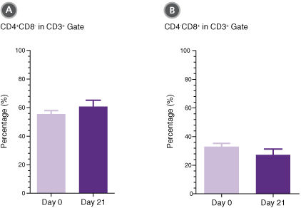
Figure 3. T Cells Expanded in ImmunoCult™-XF T Cell Expansion Medium Show Similar Proportions of CD4+ and CD8+ Cells as T Cells at the Start of Culture

T cells were isolated from human peripheral blood samples using the EasySep™ Human T Cell Isolation Kit (Catalog #17951), stimulated with ImmunoCult™ Human CD3/CD28/CD2 T Cell Activator (Catalog #10970), and cultured in ImmunoCult™-XF T Cell Expansion Medium supplemented with rhIL-2. T cells were stimulated with ImmunoCult™ Human CD3/CD28/CD2 T Cell Activator on day 0 and every 7 to 8 days for the duration of the culture. On day 0 and day 21, T cells were harvested and analyzed for (A) CD4+ and (B) CD8+ expression. Each column with error bars represents the mean ± S.E.M. (n = 24 donors for day 0 and n = 19 donors for day 21).

T Cells Expanded in ImmunoCult™-XF T Cell Expansion Medium Produce Intracellular IFN-gamma and IL-4

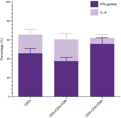
Figure 4. T Cells Expanded in ImmunoCult™-XF T Cell Expansion Medium Produce Intracellular IFN-gamma and IL-4

T cells were isolated from human peripheral blood samples using the EasySep™ Human T Cell Isolation Kit (Catalog #17951), stimulated with ImmunoCult™ Human CD3/CD28/CD2 T Cell Activator (Catalog #10970), and cultured in ImmunoCult™-XF T Cell Expansion Medium supplemented with rhIL-2. T cells were stimulated with ImmunoCult™ Human CD3/CD28/CD2 T Cell Activator on day 0 and every 7 to 8 days for the duration of the culture. On day 21, T cells were harvested and analyzed for intracellular IFN-gamma and IL-4 after stimulation with PMA and ionomycin for 4 hours and with Brefeldin A for 2 hours. The production of IFN-gamma and IL-4 in CD3+, CD3+CD4+CD8-, and CD3+CD4-CD8+ cells were determined. Each stacked column with error bars represents the mean ± S.E.M. (n = 9 donors).

Publications (8)

Cancer research 2020 sep Plasma Gelsolin Inhibits CD8+ T-cell Function and Regulates Glutathione Production to Confer Chemoresistance in Ovarian Cancer. M. Asare-Werehene et al.

Abstract

Although initial treatment of ovarian cancer is successful, tumors typically relapse and become resistant to treatment. Because of poor infiltration of effector T cells, patients are mostly unresponsive to immunotherapy. Plasma gelsolin (pGSN) is transported by exosomes (small extracellular vesicle, sEV) and plays a key role in ovarian cancer chemoresistance, yet little is known about its role in immunosurveillance. Here, we report the immunomodulatory roles of sEV-pGSN in ovarian cancer chemoresistance. In chemosensitive conditions, secretion of sEV-pGSN was low, allowing for optimal CD8+ T-cell function. This resulted in increased T-cell secretion of IFN$\gamma$, which reduced intracellular glutathione (GSH) production and sensitized chemosensitive cells to cis-diaminedichloroplatinum (CDDP)-induced apoptosis. In chemoresistant conditions, increased secretion of sEV-pGSN by ovarian cancer cells induced apoptosis in CD8+ T cells. IFN$\gamma$ secretion was therefore reduced, resulting in high GSH production and resistance to CDDP-induced death in ovarian cancer cells. These findings support our hypothesis that sEV-pGSN attenuates immunosurveillance and regulates GSH biosynthesis, a phenomenon that contributes to chemoresistance in ovarian cancer. SIGNIFICANCE: These findings provide new insight into pGSN-mediated immune cell dysfunction in ovarian cancer chemoresistance and demonstrate how this dysfunction can be exploited to enhance immunotherapy.
Cancer research 2020 oct Nano-Engineered Disruption of Heat shock protein 90 (Hsp90) Targets Drug-Induced Resistance and Relieves Natural Killer Cell Suppression in Breast Cancer. M. Smalley et al.

Abstract

Drug-induced resistance, or tolerance, is an emerging yet poorly understood failure of anticancer therapy. The interplay between drug-tolerant cancer cells and innate immunity within the tumor, the consequence on tumor growth, and therapeutic strategies to address these challenges remain undescribed. Here we elucidate the role of taxane-induced resistance on natural killer (NK) cell tumor immunity in triple-negative breast cancer (TNBC) and the design of spatio-temporally controlled nanomedicines, which boost therapeutic efficacy and invigorate 'disabled' NK. Drug tolerance limited NK cell immune surveillance via drug-induced depletion of the NK-activating ligand receptor axis, NKG2D and MHC class I polypeptide-related sequence A, B (MICA/B). Systems biology supported by empirical evidence revealed the heat shock protein 90 (Hsp90) simultaneously controls immune surveillance and persistence of drug-treated tumor cells. Based on this evidence, we engineered a 'chimeric' nano-therapeutic tool comprising taxanes and a cholesterol-tethered Hsp90 inhibitor, radicicol, which targets the tumor, reduces tolerance, and optimally re-primes NK cells via prolonged induction of NK-activating ligand receptors via temporal control of drug release in vitro and in vivo. A human ex-vivo TNBC model confirmed the importance of NK cells in drug-induced death under pressure of clinically-approved agents. These findings highlight a convergence between drug-induced resistance, the tumor-immune contexture, and engineered approaches that considers the tumor and microenvironment to improve the success of combinatorial therapy.
Nature biomedical engineering 2020 jul Classification of T-cell activation via autofluorescence lifetime imaging. A. J. Walsh et al.

Abstract

The function of a T cell depends on its subtype and activation state. Here, we show that imaging of the autofluorescence lifetime signals of quiescent and activated T cells can be used to classify the cells. T cells isolated from human peripheral blood and activated in culture using tetrameric antibodies against the surface ligands CD2, CD3 and CD28 showed specific activation-state-dependent patterns of autofluorescence lifetime. Logistic regression models and random forest models classified T cells according to activation state with 97-99{\%} accuracy, and according to activation state (quiescent or activated) and subtype (CD3+CD8+ or CD3+CD4+) with 97{\%} accuracy. Autofluorescence lifetime imaging can be used to non-destructively determine T-cell function.
Clinical cancer research : an official journal of the American Association for Cancer Research 2020 apr CUE-101, a Novel E7-pHLA-IL2-Fc Fusion Protein, Enhances Tumor Antigen-Specific T-Cell Activation for the Treatment of HPV16-Driven Malignancies. S. N. Quayle et al.

Abstract

PURPOSE To assess the potential for CUE-101, a novel therapeutic fusion protein, to selectively activate and expand HPV16 E711-20-specific CD8+ T cells as an off-the shelf therapy for the treatment of HPV16-driven tumors, including head and neck squamous cell carcinoma (HNSCC), cervical, and anal cancers. EXPERIMENTAL DESIGN CUE-101 is an Fc fusion protein composed of a human leukocyte antigen (HLA) complex, an HPV16 E7 peptide epitope, reduced affinity human IL2 molecules, and an effector attenuated human IgG1 Fc domain. Human E7-specific T cells and human peripheral blood mononuclear cells (PBMC) were tested to demonstrate cellular activity and specificity of CUE-101, whereas in vivo activity of CUE-101 was assessed in HLA-A2 transgenic mice. Antitumor efficacy with a murine surrogate (mCUE-101) was tested in the TC-1 syngeneic tumor model. RESULTS CUE-101 demonstrates selective binding, activation, and expansion of HPV16 E711-20-specific CD8+ T cells from PBMCs relative to nontarget cells. Intravenous administration of CUE-101 induced selective expansion of HPV16 E711-20-specific CD8+ T cells in HLA-A2 (AAD) transgenic mice, and anticancer efficacy and immunologic memory was demonstrated in TC-1 tumor-bearing mice treated with mCUE-101. Combination therapy with anti-PD-1 checkpoint blockade further enhanced the observed efficacy. CONCLUSIONS Consistent with its design, CUE-101 demonstrates selective expansion of an HPV16 E711-20-specific population of cytotoxic CD8+ T cells, a favorable safety profile, and in vitro and in vivo evidence supporting its potential for clinical efficacy in an ongoing phase I trial (NCT03978689).
Nature communications 2019 dec Heterogeneity and dynamics of active Kras-induced dysplastic lineages from mouse corpus stomach. J. Min et al.

Abstract

Dysplasia is considered a key transition state between pre-cancer and cancer in gastric carcinogenesis. However, the cellular or phenotypic heterogeneity and mechanisms of dysplasia progression have not been elucidated. We have established metaplastic and dysplastic organoid lines, derived from Mist1-Kras(G12D) mouse stomach corpus and studied distinct cellular behaviors and characteristics of metaplastic and dysplastic organoids. We also examined functional roles for Kras activation in dysplasia progression using Selumetinib, a MEK inhibitor, which is a downstream mediator of Kras signaling. Here, we report that dysplastic organoids die or show altered cellular behaviors and diminished aggressive behavior in response to MEK inhibition. However, the organoids surviving after MEK inhibition maintain cellular heterogeneity. Two dysplastic stem cell (DSC) populations are also identified in dysplastic cells, which exhibited different clonogenic potentials. Therefore, Kras activation controls cellular dynamics and progression to dysplasia, and DSCs might contribute to cellular heterogeneity in dysplastic cell lineages.
Nature medicine 2018 OCT Translational control of tumor immune escape via the eIF4F-STAT1-PD-L1 axis in melanoma. M. Cerezo et al.

Abstract

Preventing the immune escape of tumor cells by blocking inhibitory checkpoints, such as the interaction between programmed death ligand-1 (PD-L1) and programmed death-1 (PD-1) receptor, is a powerful anticancer approach. However, many patients do not respond to checkpoint blockade. Tumor PD-L1 expression is a potential efficacy biomarker, but the complex mechanisms underlying its regulation are not completely understood. Here, we show that the eukaryotic translation initiation complex, eIF4F, which binds the 5' cap of mRNAs, regulates the surface expression of interferon-$\gamma$-induced PD-L1 on cancer cells by regulating translation of the mRNA encoding the signal transducer and activator of transcription 1 (STAT1) transcription factor. eIF4F complex formation correlates with response to immunotherapy in human melanoma. Pharmacological inhibition of eIF4A, the RNA helicase component of eIF4F, elicits powerful antitumor immune-mediated effects via PD-L1 downregulation. Thus, eIF4A inhibitors, in development as anticancer drugs, may also act as cancer immunotherapies.
View All Publications
Top