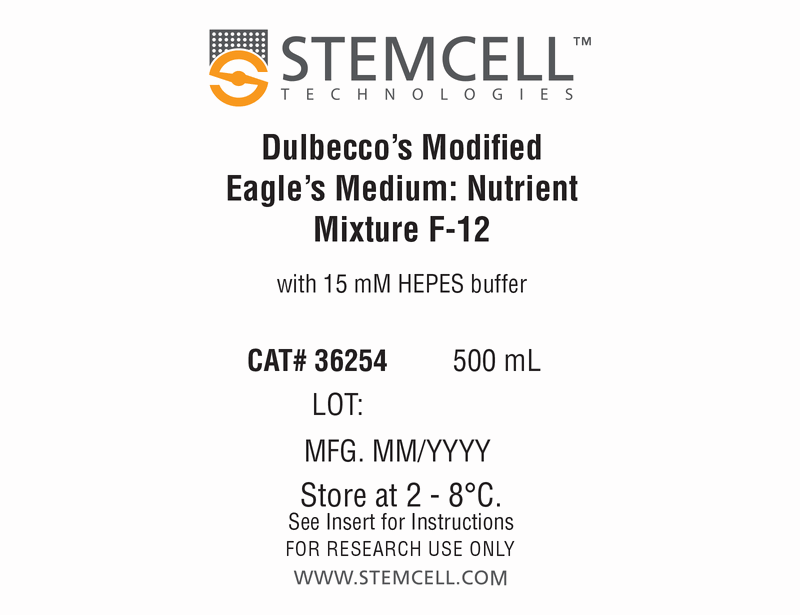
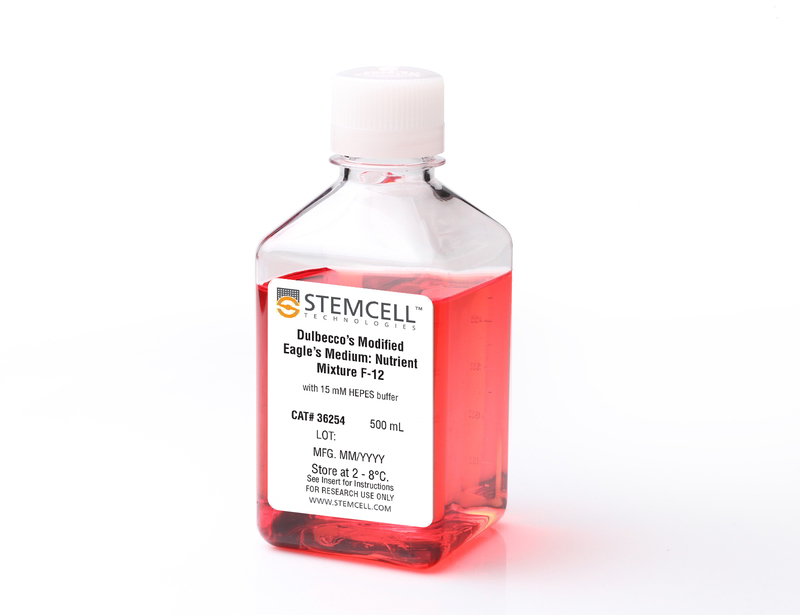
DMEM/F-12 with 15 mM HEPES
Dulbecco's Modified Eagle's Medium/Nutrient Ham's Mixture F-12 (DMEM/F-12) with 15 mM HEPES buffer
概要
Dulbecco's Modified Eagle's Medium/Nutrient Ham's Mixture F-12 (DMEM/F-12) with 15 mM HEPES is recommended for a wide variety of cell culture applications. Selection of suitable nutrient medium is dependent on cell type, culture conditions, and degree of chemical definition required for the cell culture application. This product has been pre-screened for use with other reagents of the ES-Cult™ product line for maintenance culture of embryonic stem (ES) cells in the undifferentiated state.
Subtype
Basal Media
Cell Type
Other
Species
Human, Mouse, Rat, Non-Human Primate, Other
Application
Cell Culture
技术资料
| Document Type | 产品名称 | Catalog # | Lot # | 语言 |
|---|---|---|---|---|
| Product Information Sheet | DMEM/F-12 with 15 mM HEPES | 36254 | All | English |
| Safety Data Sheet | DMEM/F-12 with 15 mM HEPES | 36254 | All | English |
数据及文献
Publications (16)
Journal of Biomedical Materials Research - Part A 2016 OCT
Efficient generation of endothelial cells from human pluripotent stem cells and characterization of their functional properties
Abstract
Abstract
Although endothelial cells (ECs) have been derived from human pluripotent stem cells (hPSCs), large-scale generation of hPSC-ECs remains challenging and their functions are not well characterized. Here we report a simple and efficient three-stage method that allows generation of approximately 98 and 9500 ECs on day 16 and day 34, respectively, from each human embryonic stem cell (hESC) input. The functional properties of hESC-ECs derived in the presence and absence of a TGF$$-inhibitory molecule SB431542 were characterized and compared with those of human umbilical vein endothelial cells (HUVECs). Confluent monolayers formed by SB431542(+) hESC-ECs, SB431542(-) hESC-ECs, and HUVECs showed similar permeability to 10,000 Da dextran, but these cells exhibited striking differences in forming tube-like structures in 3D fibrin gels. The SB431542(+) hESC-ECs were most potent in forming tube-like structures regardless of whether VEGF and bFGF were present in the medium; less potent SB431542(-) hESC-ECs and HUVECs responded differently to VEGF and bFGF, which significantly enhanced the ability of HUVECs to form tube-like structures but had little impact on SB431542(-) hESC-ECs. This study offers an efficient approach to large-scale hPSC-EC production and suggests that the phenotypes and functions of hPSC-ECs derived under different conditions need to be thoroughly examined before their use in technology development. This article is protected by copyright. All rights reserved.
International Journal of Molecular Sciences 2016 JAN
Effect of chromatin structure on the extent and distribution of DNA double strand breaks produced by ionizing radiation; comparative study of hESC and differentiated cells lines
Abstract
Abstract
Chromatin structure affects the extent of DNA damage and repair. Thus, it has been shown that heterochromatin is more protective against DNA double strand breaks (DSB) formation by ionizing radiation (IR); and that DNA DSB repair may proceed differently in hetero- and euchromatin regions. Human embryonic stem cells (hESC) have a more open chromatin structure than differentiated cells. Here, we study the effect of chromatin structure in hESC on initial DSB formation and subsequent DSB repair. DSB were scored by comet assay; and DSB repair was assessed by repair foci formation via 53BP1 antibody staining. We found that in hESC, heterochromatin is confined to distinct regions, while in differentiated cells it is distributed more evenly within the nuclei. The same dose of ionizing radiation produced considerably more DSB in hESC than in differentiated derivatives, normal human fibroblasts; and one cancer cell line. At the same time, the number of DNA repair foci were not statistically different among these cells. We showed that in hESC, DNA repair foci localized almost exclusively outside the heterochromatin regions. We also noticed that exposure to ionizing radiation resulted in an increase in heterochromatin marker H3K9me3 in cancer HT1080 cells, and to a lesser extent in IMR90 normal fibroblasts, but not in hESCs. These results demonstrate the importance of chromatin conformation for DNA protection and DNA damage repair; and indicate the difference of these processes in hESC.
Stem Cell Reviews and Reports 2016 AUG
Functionalizing Ascl1 with Novel Intracellular Protein Delivery Technology for Promoting Neuronal Differentiation of Human Induced Pluripotent Stem Cells
Abstract
Abstract
Pluripotent stem cells can become any cell type found in the body. Accordingly, one of the major challenges when working with pluripotent stem cells is producing a highly homogenous population of differentiated cells, which can then be used for downstream applications such as cell therapies or drug screening. The transcription factor Ascl1 plays a key role in neural development and previous work has shown that Ascl1 overexpression using viral vectors can reprogram fibroblasts directly into neurons. Here we report on how a recombinant version of the Ascl1 protein functionalized with intracellular protein delivery technology (Ascl1-IPTD) can be used to rapidly differentiate human induced pluripotent stem cells (hiPSCs) into neurons. We first evaluated a range of Ascl1-IPTD concentrations to determine the most effective amount for generating neurons from hiPSCs cultured in serum free media. Next, we looked at the frequency of Ascl1-IPTD supplementation in the media on differentiation and found that one time supplementation is sufficient enough to trigger the neural differentiation process. Ascl1-IPTD was efficiently taken up by the hiPSCs and enabled rapid differentiation into TUJ1-positive and NeuN-positive populations with neuronal morphology after 8 days. After 12 days of culture, hiPSC-derived neurons produced by Ascl1-IPTD treatment exhibited greater neurite length and higher numbers of branch points compared to neurons derived using a standard neural progenitor differentiation protocol. This work validates Ascl1-IPTD as a powerful tool for engineering neural tissue from pluripotent stem cells.
PLoS ONE 2015 MAR
Reprogramming of HUVECs into induced pluripotent stem cells (HiPSCs), generation and characterization of HiPSC-derived neurons and astrocytes
Abstract
Abstract
Neurodegenerative diseases are characterized by chronic and progressive structural or functional loss of neurons. Limitations related to the animal models of these human diseases have impeded the development of effective drugs. This emphasizes the need to establish disease models using human-derived cells. The discovery of induced pluripotent stem cell (iPSC) technology has provided novel opportunities in disease modeling, drug development, screening, and the potential for patient-matched" cellular therapies in neurodegenerative diseases. In this study�
Nature Communications 2015 JAN
Genetically engineering self-organization of human pluripotent stem cells into a liver bud-like tissue using Gata6
Abstract
Abstract
Human induced pluripotent stem cells (hiPSCs) have potential for personalized and regenerative medicine. While most of the methods using these cells have focused on deriving homogenous populations of specialized cells, there has been modest success in producing hiPSC-derived organotypic tissues or organoids. Here we present a novel approach for generating and then co-differentiating hiPSC-derived progenitors. With a genetically engineered pulse of GATA-binding protein 6 (GATA6) expression, we initiate rapid emergence of all three germ layers as a complex function of GATA6 expression levels and tissue context. Within 2 weeks we obtain a complex tissue that recapitulates early developmental processes and exhibits a liver bud-like phenotype, including haematopoietic and stromal cells as well as a neuronal niche. Collectively, our approach demonstrates derivation of complex tissues from hiPSCs using a single autologous hiPSCs as source and generates a range of stromal cells that co-develop with parenchymal cells to form tissues.
Nature Communications 2015 DEC
Intermediate DNA methylation is a conserved signature of genome regulation
Abstract
Abstract
The role of intermediate methylation states in DNA is unclear. Here, to comprehensively identify regions of intermediate methylation and their quantitative relationship with gene activity, we apply integrative and comparative epigenomics to 25 human primary cell and tissue samples. We report 18,452 intermediate methylation regions located near 36% of genes and enriched at enhancers, exons and DNase I hypersensitivity sites. Intermediate methylation regions average 57% methylation, are predominantly allele-independent and are conserved across individuals and between mouse and human, suggesting a conserved function. These regions have an intermediate level of active chromatin marks and their associated genes have intermediate transcriptional activity. Exonic intermediate methylation correlates with exon inclusion at a level between that of fully methylated and unmethylated exons, highlighting gene context-dependent functions. We conclude that intermediate DNA methylation is a conserved signature of gene regulation and exon usage.


