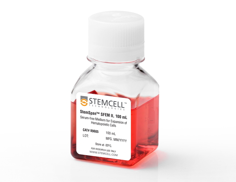
StemSpan™ SFEM II

Serum-free medium for culture and expansion of hematopoietic cells
概要
StemSpan™ Serum-Free Expansion Medium II (SFEM II) is a modified version of StemSpan™ SFEM. It has been developed for the in vitro culture and expansion of human hematopoietic cells. This medium contains pre-tested bovine serum albumin, insulin, transferrin, and other supplements in Iscove’s MDM. Recombinant hematopoietic growth factors, required for the optimal growth and expansion of hematopoietic cells, have not been added to StemSpan™ SFEM II. This allows users the flexibility to prepare medium that meets their requirements.

Using appropriate cytokines (e.g. StemSpan™ CC100, StemSpan™ CC110, or StemSpan™ CD34+ Expansion Supplement), StemSpan™ SFEM II can be used for the expansion of total nucleated cells and CD34+ cells from cord blood, bone marrow, or other cell sources. StemSpan™ SFEM II can also be used to expand and differentiate lineage-committed progenitor cells to generate erythroblasts, granulocytes, monocytes, or megakaryocytes when used with StemSpan™ Erythroid Expansion Supplement (Catalog #02692), StemSpan™ Myeloid Expansion Supplement (Catalog #02693), StemSpan™ Myeloid Expansion Supplement II (Catalog #02694), or StemSpan™ Megakaryocyte Expansion Supplement (Catalog #02696), respectively.
Contains
• Iscove’s MDM
• Bovine serum albumin
• Recombinant human insulin
• Human transferrin (iron-saturated)
• 2-Mercaptoethanol
• Supplements
Subtype
Specialized Media
Cell Type
Hematopoietic Stem and Progenitor Cells
Species
Human
Application
Cell Culture, Expansion
Brand
StemSpan
Area of Interest
Stem Cell Biology, Transplantation Research
Formulation
Serum-Free
技术资料
Document Type 产品名称 Catalog # Lot # 语言
Product Information Sheet StemSpan™ SFEM II 09605, 09655 All English
Safety Data Sheet StemSpan™ SFEM II 09605, 09655 All English
数据及文献

Data

Expansion of CD34 + Human Cord Blood Cells Cultured in StemSpan™ Media Containing CC100 Cytokine Cocktail

Figure 1. Expansion of CD34 + Human Cord Blood Cells Cultured in StemSpan™ Media Containing CC100 Cytokine Cocktail

Purified CD34 + human cord blood (CB) cells were suspended at a concentration of 10,000 per mL in StemSpan™ SFEM (dark gray bars), SFEM II (gold bars) and ACF (orange bars) media containing CC100 Cytokine Cocktail (Catalog #02690). Cultures were maintained for 7 days, after which the cells were counted and examined for CD34 and CD45 expression by flow cytometry. Shown are the fold expansion of total nucleated cells (TNC) (A) and CD34 + cells (B) per input CD34 + cell, and the percent CD34 + cells (C). Results represent the average results of 32 different CB samples. Vertical lines indicate 95% confidence limits, the range within which 95% of results fall. The numbers of cells produced in StemSpan™ SFEM II were significantly higher than in StemSpan™ SFEM and StemSpan™-ACF (*p<0.001, paired t-test, n=32).

StemSpan™ SFEM II Serum-Free Expansion Medium Containing CC100 Cytokine Cocktail Supports Greater Expansion of Human CD34 + Cells Than Other Media Tested

Figure 2. StemSpan™ SFEM II Serum-Free Expansion Medium Containing CC100 Cytokine Cocktail Supports Greater Expansion of Human CD34 + Cells Than Other Media Tested

Expansion of CD34 + cells, normalized relative to the values obtained in StemSpan™ SFEM medium (dark gray bars) after culturing purified CD34 + CB (A, n=6) or bone marrow (BM) (B, n=3) cells for 7 days in StemSpan™ SFEM, SFEM II (gold bars) and ACF (orange bars), and six media from other commercial suppliers (light gray bars, Competitor 1-6, which included, in random order, StemPro34 (Life Technologies), X-Vivo-15 and HPGM (both from Lonza), SCGM (Cellgenix), StemLine II (Sigma) and HP01 (Macopharma)). All media were supplemented with StemSpan™ CC100 Cytokine Cocktail (Catalog #02690). Vertical lines indicate 95% confidence limits, the range within which 95% of results fall. The numbers of CB and BM cells produced in StemSpan™ SFEM II were significantly higher than in all other media, except the numbers of CB cells produced in StemSpan™-ACF (*p<0.05, paired t-test).

Expansion of CD34 + Human Cord Blood Cells Cultured in StemSpan™ Media Containing CD34 + Expansion Supplement

Figure 3. Expansion of CD34 + Human Cord Blood Cells Cultured in StemSpan™ Media Containing CD34 + Expansion Supplement

Purified CD34 + human cord blood (CB) cells were suspended at a concentration of 10,000 per mL in StemSpan™ SFEM (dark gray bars), SFEM II (gold bars) and ACF (orange bars) media containing CD34 + Expansion Supplement (Catalog #02691). Cultures were maintained for 7 days, after which the cells were counted and examined for CD34 and CD45 expression by flow cytometry. The number of colony-forming units (CFU) in the expanded population was determined by replating cells in MethoCult™ H4435 and counting the number of colonies produced 14 days later. Shown are the fold expansion of total nucleated cells (TNC) (A), CD34 + cells (B) and CFU numbers (C) per input CD34 + cell, and the percent CD34 + cells (D) in these cultures (n=6). Vertical lines indicate 95% confidence limits, the range within which 95% of results fall. The numbers of cells produced in StemSpan™ SFEM II was significantly higher than in SFEM and ACF (*p<0.001, #p<0.05, paired t-test, n=6).

StemSpan™ SFEM II Serum-Free Expansion Medium Containing CD34 + Expansion Supplement Supports Greater Expansion of Human CD34 + Cells Than Other Media Tested

Figure 4. StemSpan™ SFEM II Serum-Free Expansion Medium Containing CD34 + Expansion Supplement Supports Greater Expansion of Human CD34 + Cells Than Other Media Tested

Expansion of CD34 + cells (A) and CFUs (B), normalized relative to the values obtained in SFEM medium (dark gray bars) after culturing purified CD34 + CB cells for 7 days in StemSpan™ SFEM, SFEM II (gold bars) and ACF (orange bars), and six media from other suppliers (light gray bars, Competitor 1-6, which included, in random order, X-Vivo-15 (Lonza), HP01 (Macopharma), StemPro34 (Life Technologies), SCGM (Cellgenix), StemLine II (Sigma), and HPGM (Lonza). All media were supplemented with the StemSpan™ CD34 + Expansion Supplement (Catalog #02691). Vertical lines indicate 95% confidence limits, the range within which 95% of results fall. The numbers of cells produced in StemSpan™ SFEM II were significantly higher than in all other media (*p<0.01, paired t-test, n=6).

StemSpan™ SFEM II Serum-Free Expansion Medium Containing Erythroid Expansion Supplement Supports Greater Expansion of Erythroid Cells Than Other Media Tested

Figure 5. StemSpan™ SFEM II Serum-Free Expansion Medium Containing Erythroid Expansion Supplement Supports Greater Expansion of Erythroid Cells Than Other Media Tested

The numbers of erythroid cells, normalized relative to the values obtained in StemSpan™ SFEM medium (dark gray bar), obtained after culturing purified CD34 + CB cells for 14 days in StemSpan™ SFEM, SFEM II (gold bars) and ACF (orange bars), and six media from other commercial suppliers (light gray bars, Competitor 1-6, which included, in random order, X-Vivo-15 and HPGM (both from Lonza), StemLine II (Sigma), HP01 (Macopharma), StemPro34 (Life Technologies) and SCGM (Cellgenix). All media were supplemented with StemSpan™ Erythroid Expansion Supplement (Catalog #02692). Vertical lines indicate 95% confidence limits, the range within which 95% of results fall. The numbers of cells produced in StemSpan™ SFEM II were significantly higher than in all other media (*p<0.05, paired t-test, n=6).

Table 1. Production of Myeloid Cells from Human CB CD34+ Cells Cultured in SFEM II Containing Myeloid Expansion Supplement or Myeloid Expansion Supplement ll

StemSpan™ SFEM II Serum-Free Expansion Medium Containing Erythroid Expansion Supplement Supports Greater Expansion of Erythroid Cells Than Other Media Tested

Shown are numbers of total nucleated cells (TNCs) produced per input human CB-derived CD34+ cell and percentages of cells positive for myeloid markers CD13, CD14 and CD15 after 14 days of culture in SFEM II containing Myeloid Expansion Supplement (n = 14) or Myeloid Expansion Supplement II (n = 16). *95% confidence limits (CL); the range within which 95% of results typically fall.

StemSpan™ SFEM II Serum-Free Expansion Medium Containing Megakaryocyte Expansion Supplement Supports Greater Expansion of Megakaryocytes Than Other Media Tested

Figure 6. StemSpan™ SFEM II Serum-Free Expansion Medium Containing Megakaryocyte Expansion Supplement Supports Greater Expansion of Megakaryocytes Than Other Media Tested

The numbers of megakaryocytes, normalized relative to the values obtained in StemSpan™ SFEM medium (dark gray bar), obtained after culturing purified CD34 + CB cells for 14 days in StemSpan™ SFEM, SFEM II (gold bars) and ACF (orange bars), and six media from other commercial suppliers (light gray bars, Competitor 1-6, which included, in random order, StemLine II (Sigma), HPGM (Lonza), HP01 (Macopharma), SCGM (Cellgenix), StemPro34 (Life Technologies) and X-Vivo-15 (Lonza). All media were supplemented with StemSpan™ Megakaryocyte Expansion Supplement (Catalog #02696). Vertical lines indicate 95% confidence limits, the range within which 95% of results fall. The numbers of cells produced in the StemSpan™ media were significantly higher than in the other media (*p<0.01 paired t-test, n=6).
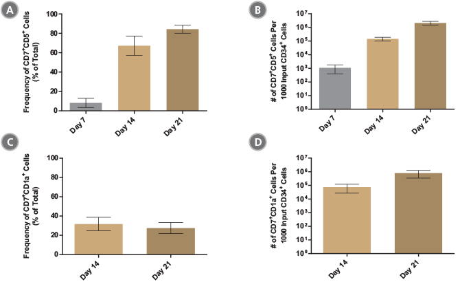
Figure 7. StemSpan™ SFEM II Serum-Free Expansion Medium Containing T Cell Progenitor Expansion Supplement Promotes the Expansion and Differentiation of CB-Derived CD34+ Cells into Pro- and Pre-T Cells

The average (A,C) frequencies and (B,D) numbers of (A,B) CD7+CD5+ pro-T cells and (C,D) CD7+CD1a+ pre-T cells on days 7, 14 and 21 of culture with the StemSpan™ T Cell Progenitor Differentiation Kit (Catalog #09900) are shown for 10 - 26 independent experiments. The average frequency of (A) pro- and (C) pre-T cells were 84% and 28% respectively, after 21 days of culture. All pro- and pre-T cells were found to express intracellular CD3 (data not shown). The number of (B) CD7+CD5+ pro-T cells increased (on average) ~10 - 100-fold every week, resulting in an average number of ~2100 pro-T cells produced per input CD34+ cell on day 21. After 21 days of culture (D) pre-T cells expressing CD7 and CD1a are present in large numbers, indicating the further differentiation of pro-T cells. The yield of (D) CD7+CD1a+ cells on day 21 was ~800 per input CD34+ cell. BM-derived CD34+ cells also expanded and differentiated to pro-T cells in stroma-free cultures with approximately 70 CD7+CD5+ cells produced per input CD34+ cell at day 21 (n = 3; data not shown). Vertical lines indicate 95% confidence limits (CL), the range within which 95% of results typically fall.

Publications (14)

Nature communications 2019 sep Tissue-resident memory CD8+ T cells amplify anti-tumor immunity by triggering antigen spreading through dendritic cells. E. Menares et al.

Abstract

Tissue-resident memory CD8+ T (Trm) cells mediate potent local innate and adaptive immune responses and play a central role against solid tumors. However, whether Trm cells cross-talk with dendritic cells (DCs) to support anti-tumor immunity remains unclear. Here we show that antigen-specific activation of skin Trm cells leads to maturation and migration to draining lymph nodes of cross-presenting dermal DCs. Tumor rejection mediated by Trm cells triggers the spread of cytotoxic CD8+ T cell responses against tumor-derived neo- and self-antigens via dermal DCs. These responses suppress the growth of intradermal tumors and disseminated melanoma lacking the Trm cell-targeted epitope. Moreover, analysis of RNA sequencing data from human melanoma tumors reveals that enrichment of a Trm cell gene signature associates with DC activation and improved survival. This work unveils the ability of Trm cells to amplify the breath of cytotoxic CD8+ T cell responses through DCs, thereby strengthening anti-tumor immunity.
Cell stem cell 2019 mar The NAD-Booster Nicotinamide Riboside Potently Stimulates Hematopoiesis through Increased Mitochondrial Clearance. N. Vannini et al.

Abstract

It has been recently shown that increased oxidative phosphorylation, as reflected by increased mitochondrial activity, together with impairment of the mitochondrial stress response, can severely compromise hematopoietic stem cell (HSC) regeneration. Here we show that the NAD+-boosting agent nicotinamide riboside (NR) reduces mitochondrial activity within HSCs through increased mitochondrial clearance, leading to increased asymmetric HSC divisions. NR dietary supplementation results in a significantly enlarged pool of progenitors, without concurrent HSC exhaustion, improves survival by 80{\%}, and accelerates blood recovery after murine lethal irradiation and limiting-HSC transplantation. In immune-deficient mice, NR increased the production of human leucocytes from hCD34+ progenitors. Our work demonstrates for the first time a positive effect of NAD+-boosting strategies on the most primitive blood stem cells, establishing a link between HSC mitochondrial stress, mitophagy, and stem-cell fate decision, and unveiling the potential of NR to improve recovery of patients suffering from hematological failure including post chemo- and radiotherapy.
Science translational medicine 2019 jul Therapeutically relevant engraftment of a CRISPR-Cas9-edited HSC-enriched population with HbF reactivation in nonhuman primates. O. Humbert et al.

Abstract

Reactivation of fetal hemoglobin (HbF) is being pursued as a treatment strategy for hemoglobinopathies. Here, we evaluated the therapeutic potential of hematopoietic stem and progenitor cells (HSPCs) edited with the CRISPR-Cas9 nuclease platform to recapitulate naturally occurring mutations identified in individuals who express increased amounts of HbF, a condition known as hereditary persistence of HbF. CRISPR-Cas9 treatment and transplantation of HSPCs purified on the basis of surface expression of the CD34 receptor in a nonhuman primate (NHP) autologous transplantation model resulted in up to 30{\%} engraftment of gene-edited cells for >1 year. Edited cells effectively and stably reactivated HbF, as evidenced by up to 18{\%} HbF-expressing erythrocytes in peripheral blood. Similar results were obtained by editing highly enriched stem cells, defined by the markers CD34+CD90+CD45RA-, allowing for a 10-fold reduction in the number of transplanted target cells, thus considerably reducing the need for editing reagents. The frequency of engrafted, gene-edited cells persisting in vivo using this approach may be sufficient to ameliorate the phenotype for a number of genetic diseases.
Cell stem cell 2019 aug Interconversion between Tumorigenic and Differentiated States in Acute Myeloid Leukemia. M. D. McKenzie et al.

Abstract

Tumors are composed of phenotypically heterogeneous cancer cells that often resemble various differentiation states of their lineage of origin. Within this hierarchy, it is thought that an immature subpopulation of tumor-propagating cancer stem cells (CSCs) differentiates into non-tumorigenic progeny, providing a rationale for therapeutic strategies that specifically eradicate CSCs or induce their differentiation. The clinical success of these approaches depends on CSC differentiation being unidirectional rather than reversible, yet this question remains unresolved even in prototypically hierarchical malignancies, such as acute myeloid leukemia (AML). Here, we show in murine and human models of AML that, upon perturbation of endogenous expression of the lineage-determining transcription factor PU.1 or withdrawal of established differentiation therapies, some mature leukemia cells can de-differentiate and reacquire clonogenic and leukemogenic properties. Our results reveal plasticity of CSC maturation in AML, highlighting the need to therapeutically eradicate cancer cells across a range of differentiation states.
Nature communications 2019 Gene correction for SCID-X1 in long-term hematopoietic stem cells. M. Pavel-Dinu et al.

Abstract

Gene correction in human long-term hematopoietic stem cells (LT-HSCs) could be an effective therapy for monogenic diseases of the blood and immune system. Here we describe an approach for X-linked sSevere cCombined iImmunodeficiency (SCID-X1) using targeted integration of a cDNA into the endogenous start codon to functionally correct disease-causing mutations throughout the gene. Using a CRISPR-Cas9/AAV6 based strategy, we achieve up to 20{\%} targeted integration frequencies in LT-HSCs. As measures of the lack of toxicity we observe no evidence of abnormal hematopoiesis following transplantation and no evidence of off-target mutations using a high-fidelity Cas9 as a ribonucleoprotein complex. We achieve high levels of targeting frequencies (median 45{\%}) in CD34+ HSPCs from six SCID-X1 patients and demonstrate rescue of lymphopoietic defect in a patient derived HSPC population in vitro and in vivo. In sum, our study provides specificity, toxicity and efficacy data supportive of clinical development of genome editing to treat SCID-Xl.
Molecular therapy. Nucleic acids 2018 SEP Priming Human Repopulating Hematopoietic Stem and Progenitor Cells for Cas9/sgRNA Gene Targeting. C. T. Charlesworth et al.

Abstract

Engineered nuclease-mediated gene targeting through homologous recombination (HR) in hematopoietic stem and progenitor cells (HSPCs) has the potential to treat a variety of genetic hematologic and immunologic disorders. Here, we identify critical parameters to reproducibly achieve high frequencies of RNA-guided (single-guide RNA [sgRNA]; CRISPR)-Cas9 nuclease (Cas9/sgRNA) and rAAV6-mediated HR at the $\beta$-globin (HBB) locus in HSPCs. We identified that by transducing HSPCs with rAAV6 post-electroporation, there was a greater than 2-fold electroporation-aided transduction (EAT) of rAAV6 endocytosis with roughly 70{\%} of the cell population having undergone transduction within 2 hr. When HSPCs are cultured at low densities (1 × 10⁵ cells/mL) prior to HBB targeting, HSPC expansion rates are significantly positively correlated with HR frequencies in vitro as well as in repopulating cells in immunodeficient NSG mice in vivo. We also show that culturing fluorescence-activated cell sorting (FACS)-enriched HBB-targeted HSPCs at low cell densities in the presence of the small molecules, UM171 and SR1, stimulates the expansion of gene-edited HSPCs as measured by higher engraftment levels in immunodeficient mice. This work serves not only as an optimized protocol for genome editing HSPCs at the HBB locus for the treatment of $\beta$-hemoglobinopathies but also as a foundation for editing HSPCs at other loci for both basic and translational research.
View All Publications
Top