StemSpan™ SFEM

Serum-free medium for culture and expansion of hematopoietic cells
概要
StemSpan™ Serum-Free Expansion Medium (SFEM) has been developed and tested for the in vitro culture and expansion of human hematopoietic cells, when the appropriate growth factors and supplements are added. This allows users the flexibility to prepare medium that meets their requirements. When combined with the appropriate cytokines, SFEM has been used for the culture and expansion of hematopoietic cells isolated from other species, including mouse, non-human primate, and dog. SFEM has also been used for culture of various other hematopoietic and non-hematopoietic cell types. Using appropriate StemSpan™ Expansion Supplements, SFEM may be used to expand CD34+ cells isolated from human cord blood, mobilized peripheral blood, or bone marrow samples, or to expand and differentiate lineage-committed progenitors to generate populations of erythroid, myeloid, or megakaryocyte progenitor cells.

StemSpan™ SFEM II (Catalog #09605) is an improved version of StemSpan™ SFEM that is further enriched to promote and support higher rates of CD34+ expansion and/or cell differentiation.
Contains
• Iscove’s MDM
• Bovine serum albumin
• Recombinant human insulin
• Human transferrin (iron-saturated)
• 2-Mercaptoethanol
• Supplements
Subtype
Specialized Media
Cell Type
Hematopoietic Stem and Progenitor Cells
Species
Human, Mouse, Rat, Non-Human Primate
Application
Cell Culture, Expansion
Brand
StemSpan
Area of Interest
Stem Cell Biology, Transplantation Research
Formulation
Serum-Free
技术资料
Document Type 产品名称 Catalog # Lot # 语言
Product Information Sheet StemSpan™ SFEM 09600, 09650 All English
Safety Data Sheet StemSpan™ SFEM 09600, 09650 All English
数据及文献

Data

Expansion of CD34 + Human Cord Blood Cells Cultured in StemSpan™ Media Containing CC100 Cytokine Cocktail

Figure 1. Expansion of CD34 + Human Cord Blood Cells Cultured in StemSpan™ Media Containing CC100 Cytokine Cocktail

Purified CD34 + human cord blood (CB) cells were suspended at a concentration of 10,000 per mL in StemSpan™ SFEM (dark gray bars), SFEM II (gold bars) and ACF (orange bars) media containing CC100 Cytokine Cocktail (Catalog #02690). Cultures were maintained for 7 days, after which the cells were counted and examined for CD34 and CD45 expression by flow cytometry. Shown are the fold expansion of total nucleated cells (TNC) (A) and CD34 + cells (B) per input CD34 + cell, and the percent CD34 + cells (C). Results represent the average results of 32 different CB samples. Vertical lines indicate 95% confidence limits, the range within which 95% of results fall. The numbers of cells produced in StemSpan™ SFEM II were significantly higher than in StemSpan™ SFEM and StemSpan™-ACF (*p<0.001, paired t-test, n=32).

StemSpan™ SFEM II Serum-Free Expansion Medium Containing CC100 Cytokine Cocktail Supports Greater Expansion of Human CD34 + Cells Than Other Media Tested

Figure 2. StemSpan™ SFEM II Serum-Free Expansion Medium Containing CC100 Cytokine Cocktail Supports Greater Expansion of Human CD34 + Cells Than Other Media Tested

Expansion of CD34 + cells, normalized relative to the values obtained in StemSpan™ SFEM medium (dark gray bars) after culturing purified CD34 + CB (A, n=6) or bone marrow (BM) (B, n=3) cells for 7 days in StemSpan™ SFEM, SFEM II (gold bars) and ACF (orange bars), and six media from other commercial suppliers (light gray bars, Competitor 1-6, which included, in random order, StemPro34 (Life Technologies), X-Vivo-15 and HPGM (both from Lonza), SCGM (Cellgenix), StemLine II (Sigma) and HP01 (Macopharma)). All media were supplemented with StemSpan™ CC100 Cytokine Cocktail (Catalog #02690). Vertical lines indicate 95% confidence limits, the range within which 95% of results fall. The numbers of CB and BM cells produced in StemSpan™ SFEM II were significantly higher than in all other media, except the numbers of CB cells produced in StemSpan™-ACF (*p<0.05, paired t-test).

Expansion of CD34 + Human Cord Blood Cells Cultured in StemSpan™ Media Containing CD34 + Expansion Supplement

Figure 3. Expansion of CD34 + Human Cord Blood Cells Cultured in StemSpan™ Media Containing CD34 + Expansion Supplement

Purified CD34 + human cord blood (CB) cells were suspended at a concentration of 10,000 per mL in StemSpan™ SFEM (dark gray bars), SFEM II (gold bars) and ACF (orange bars) media containing CD34 + Expansion Supplement (Catalog #02691). Cultures were maintained for 7 days, after which the cells were counted and examined for CD34 and CD45 expression by flow cytometry. The number of colony-forming units (CFU) in the expanded population was determined by replating cells in MethoCult™ H4435 and counting the number of colonies produced 14 days later. Shown are the fold expansion of total nucleated cells (TNC) (A), CD34 + cells (B) and CFU numbers (C) per input CD34 + cell, and the percent CD34 + cells (D) in these cultures (n=6). Vertical lines indicate 95% confidence limits, the range within which 95% of results fall. The numbers of cells produced in StemSpan™ SFEM II was significantly higher than in SFEM and ACF (*p<0.001, #p<0.05, paired t-test, n=6).

StemSpan™ SFEM II Serum-Free Expansion Medium Containing CD34 + Expansion Supplement Supports Greater Expansion of Human CD34 + Cells Than Other Media Tested

Figure 4. StemSpan™ SFEM II Serum-Free Expansion Medium Containing CD34 + Expansion Supplement Supports Greater Expansion of Human CD34 + Cells Than Other Media Tested

Expansion of CD34 + cells (A) and CFUs (B), normalized relative to the values obtained in SFEM medium (dark gray bars) after culturing purified CD34 + CB cells for 7 days in StemSpan™ SFEM, SFEM II (gold bars) and ACF (orange bars), and six media from other suppliers (light gray bars, Competitor 1-6, which included, in random order, X-Vivo-15 (Lonza), HP01 (Macopharma), StemPro34 (Life Technologies), SCGM (Cellgenix), StemLine II (Sigma), and HPGM (Lonza). All media were supplemented with the StemSpan™ CD34 + Expansion Supplement (Catalog #02691). Vertical lines indicate 95% confidence limits, the range within which 95% of results fall. The numbers of cells produced in StemSpan™ SFEM II were significantly higher than in all other media (*p<0.01, paired t-test, n=6).

Table 1. Production of Erythroid Cells From CD34 + Human Cord Blood Cells Cultured in StemSpan™ SFEM Serum-Free Expansion Medium Containing Erythroid Expansion Supplement

Production of Erythroid Cells From CD34 + Human Cord Blood Cells Cultured in StemSpan™ SFEM Serum-Free Expansion Medium Containing Erythroid Expansion Supplement

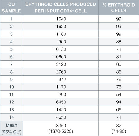
Numbers and percent of erythroid cells produced after 14 days of culture of enriched CD34 + cells from 14 different cord blood (CB) samples. Erythroid cells were characterized by flow cytometry on the basis of transferrin receptor (CD71) and glycophorin A (CD235) expression.*95% confidence limits, the range within which 95% of the results fall.

StemSpan™ SFEM II Serum-Free Expansion Medium Containing Erythroid Expansion Supplement Supports Greater Expansion of Erythroid Cells Than Other Media Tested

Figure 5. StemSpan™ SFEM II Serum-Free Expansion Medium Containing Erythroid Expansion Supplement Supports Greater Expansion of Erythroid Cells Than Other Media Tested

The numbers of erythroid cells, normalized relative to the values obtained in StemSpan™ SFEM medium (dark gray bar), obtained after culturing purified CD34 + CB cells for 14 days in StemSpan™ SFEM, SFEM II (gold bars) and ACF (orange bars), and six media from other commercial suppliers (light gray bars, Competitor 1-6, which included, in random order, X-Vivo-15 and HPGM (both from Lonza), StemLine II (Sigma), HP01 (Macopharma), StemPro34 (Life Technologies) and SCGM (Cellgenix). All media were supplemented with StemSpan™ Erythroid Expansion Supplement (Catalog #02692). Vertical lines indicate 95% confidence limits, the range within which 95% of results fall. The numbers of cells produced in StemSpan™ SFEM II were significantly higher than in all other media (*p<0.05, paired t-test, n=6).

Table 2. Production of Megakaryocytes From CD34+ Human Cord Blood Cells Cultured in StemSpan™ SFEM Serum-Free Expansion Medium Containing Megakaryocyte Expansion Supplement

Production of Megakaryocytes From CD34+ Human Cord Blood Cells Cultured in StemSpan™ SFEM Serum-Free Expansion Medium Containing Megakaryocyte Expansion Supplement

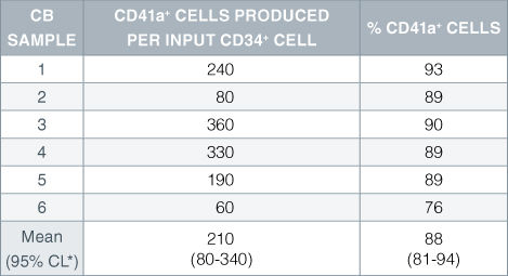
Numbers and percent of cells expressing the megakaryocyte marker CD41a produced after 14 days of culture of enriched CD34 + cells from 6 independent cord blood (CB) samples. *95% confidence limits, the range within which 95% of the results fall.

StemSpan™ SFEM II Serum-Free Expansion Medium Containing Megakaryocyte Expansion Supplement Supports Greater Expansion of Megakaryocytes Than Other Media Tested

Figure 6. StemSpan™ SFEM II Serum-Free Expansion Medium Containing Megakaryocyte Expansion Supplement Supports Greater Expansion of Megakaryocytes Than Other Media Tested

The numbers of megakaryocytes, normalized relative to the values obtained in StemSpan™ SFEM medium (dark gray bar), obtained after culturing purified CD34 + CB cells for 14 days in StemSpan™ SFEM, SFEM II (gold bars) and ACF (orange bars), and six media from other commercial suppliers (light gray bars, Competitor 1-6, which included, in random order, StemLine II (Sigma), HPGM (Lonza), HP01 (Macopharma), SCGM (Cellgenix), StemPro34 (Life Technologies) and X-Vivo-15 (Lonza). All media were supplemented with StemSpan™ Megakaryocyte Expansion Supplement (Catalog #02696). Vertical lines indicate 95% confidence limits, the range within which 95% of results fall. The numbers of cells produced in the StemSpan™ media were significantly higher than in the other media (*p<0.01 paired t-test, n=6).

Publications (223)

Cell stem cell 2020 Restraining Lysosomal Activity Preserves Hematopoietic Stem Cell Quiescence and Potency. R. Liang et al.

Abstract

Quiescence is a fundamental property that maintains hematopoietic stem cell (HSC) potency throughout life. Quiescent HSCs are thought to rely on glycolysis for their energy, but the overall metabolic properties of HSCs remain elusive. Using combined approaches, including single-cell RNA sequencing (RNA-seq), we show that mitochondrial membrane potential (MMP) distinguishes quiescent from cycling-primed HSCs. We found that primed, but not quiescent, HSCs relied readily on glycolysis. Notably, in vivo inhibition of glycolysis enhanced the competitive repopulation ability of primed HSCs. We further show that HSC quiescence is maintained by an abundance of large lysosomes. Repression of lysosomal activation in HSCs led to further enlargement of lysosomes while suppressing glucose uptake. This also induced increased lysosomal sequestration of mitochondria and enhanced the competitive repopulation ability of primed HSCs by over 90-fold in vivo. These findings show that restraining lysosomal activity preserves HSC quiescence and potency and may be therapeutically relevant.
PloS one 2020 A profile of circulating vascular progenitor cells in human neovascular age-related macular degeneration. T. Catchpole et al.

Abstract

BACKGROUND/OBJECTIVE A subset of neovascular age-related macular degeneration (nvAMD) subjects appears to be refractory to the effects of anti-VEGF treatment and require frequent intravitreal injections. The vascular phenotype of the choroidal neovascular (CNV) lesions may contribute to the resistance. Animal studies of CNV lesions have shown that cells originating from bone marrow are capable of forming varying cell types in the lesions. This raised the possibility of a similar cell population in human nvAMD subjects. MATERIALS AND METHODS Blood draws were obtained from subjects with active nvAMD while patients were receiving standard of care anti-VEGF injections. Subjects were classified as refractory or non-refractory to anti-VEGF treatment based on previous number of injections in the preceding 12 months. Peripheral blood mononuclear cells (PBMCs) were isolated and CD34-positive cells purified using magnetic bead sorting. The isolated cells were expanded in StemSpan SFEM media to increase cell numbers. After expansion, the cells were split and plated in either endothelial or mesenchymal promoting conditions. Phenotype analysis was performed via qPCR. RESULTS There was no significant difference in the number of PBMCs and CD34-positive cells between refractory and non-refractory nvAMD subjects. The growth pattern distribution between endothelial and mesenchymal media conditions were very similar between refractory and non-refractory subjects. qPCR and immunostaining demonstrated positive expression of endothelial markers in endothelial media, and markers such as NG2 and $\alpha$SMA in mesenchymal media. However, analysis of subsequent samples from AMD subjects demonstrated high variability in both the numbers and differentiation properties of this cell population. CONCLUSIONS CD34+ cells can be isolated from nvAMD subjects and show both endothelial and pericyte-like characteristics after differentiation in certain media conditions. However, nvAMD subjects show high variability in both numbers of cells and differentiation characteristics in repeat sampling. This variability highlights the importance of taking multiple samples from nvAMD subjects for any clinical trials focused on biomarkers for the disease.
Nature materials 2019 may Targeted homology-directed repair in blood stem and progenitor cells with CRISPR nanoformulations. R. Shahbazi et al.

Abstract

Ex vivo CRISPR gene editing in haematopoietic stem and progenitor cells has opened potential treatment modalities for numerous diseases. The current process uses electroporation, sometimes followed by virus transduction. While this complex manipulation has resulted in high levels of gene editing at some genetic loci, cellular toxicity was observed. We have developed a CRISPR nanoformulation based on colloidal gold nanoparticles with a unique loading design capable of cellular entry without the need for electroporation or viruses. This highly monodispersed nanoformulation avoids lysosomal entrapment and localizes to the nucleus in primary human blood progenitors without toxicity. Nanoformulation-mediated gene editing is efficient and sustained with different CRISPR nucleases at multiple loci of therapeutic interest. The engraftment kinetics of nanoformulation-treated primary cells in humanized mice are better relative to those of non-treated cells, with no differences in differentiation. Here we demonstrate non-toxic delivery of the entire CRISPR payload into primary human blood progenitors.
Cell stem cell 2019 mar The NAD-Booster Nicotinamide Riboside Potently Stimulates Hematopoiesis through Increased Mitochondrial Clearance. N. Vannini et al.

Abstract

It has been recently shown that increased oxidative phosphorylation, as reflected by increased mitochondrial activity, together with impairment of the mitochondrial stress response, can severely compromise hematopoietic stem cell (HSC) regeneration. Here we show that the NAD+-boosting agent nicotinamide riboside (NR) reduces mitochondrial activity within HSCs through increased mitochondrial clearance, leading to increased asymmetric HSC divisions. NR dietary supplementation results in a significantly enlarged pool of progenitors, without concurrent HSC exhaustion, improves survival by 80{\%}, and accelerates blood recovery after murine lethal irradiation and limiting-HSC transplantation. In immune-deficient mice, NR increased the production of human leucocytes from hCD34+ progenitors. Our work demonstrates for the first time a positive effect of NAD+-boosting strategies on the most primitive blood stem cells, establishing a link between HSC mitochondrial stress, mitophagy, and stem-cell fate decision, and unveiling the potential of NR to improve recovery of patients suffering from hematological failure including post chemo- and radiotherapy.
Stem cells (Dayton, Ohio) 2019 jul p53-TP53-Induced Glycolysis Regulator Mediated Glycolytic Suppression Attenuates DNA Damage and Genomic Instability in Fanconi Anemia Hematopoietic Stem Cells. X. Li et al.

Abstract

Emerging evidence has shown that resting quiescent hematopoietic stem cells (HSCs) prefer to utilize anaerobic glycolysis rather than mitochondrial respiration for energy production. Compelling evidence has also revealed that altered metabolic energetics in HSCs underlies the onset of certain blood diseases; however, the mechanisms responsible for energetic reprogramming remain elusive. We recently found that Fanconi anemia (FA) HSCs in their resting state are more dependent on mitochondrial respiration for energy metabolism than on glycolysis. In the present study, we investigated the role of deficient glycolysis in FA HSC maintenance. We observed significantly reduced glucose consumption, lactate production, and ATP production in HSCs but not in the less primitive multipotent progenitors or restricted hematopoietic progenitors of Fanca-/- and Fancc-/- mice compared with that of wild-type mice, which was associated with an overactivated p53 and TP53-induced glycolysis regulator, the TIGAR-mediated metabolic axis. We utilized Fanca-/- HSCs deficient for p53 to show that the p53-TIGAR axis suppressed glycolysis in FA HSCs, leading to enhanced pentose phosphate pathway and cellular antioxidant function and, consequently, reduced DNA damage and attenuated HSC exhaustion. Furthermore, by using Fanca-/- HSCs carrying the separation-of-function mutant p53R172P transgene that selectively impairs the p53 function in apoptosis but not cell-cycle control, we demonstrated that the cell-cycle function of p53 was not required for glycolytic suppression in FA HSCs. Finally, ectopic expression of the glycolytic rate-limiting enzyme PFKFB3 specifically antagonized p53-TIGAR-mediated metabolic reprogramming in FA HSCs. Together, our results suggest that p53-TIGAR metabolic axis-mediated glycolytic suppression may play a compensatory role in attenuating DNA damage and proliferative exhaustion in FA HSCs. Stem Cells 2019;37:937-947.
Cell stem cell 2019 aug Interconversion between Tumorigenic and Differentiated States in Acute Myeloid Leukemia. M. D. McKenzie et al.

Abstract

Tumors are composed of phenotypically heterogeneous cancer cells that often resemble various differentiation states of their lineage of origin. Within this hierarchy, it is thought that an immature subpopulation of tumor-propagating cancer stem cells (CSCs) differentiates into non-tumorigenic progeny, providing a rationale for therapeutic strategies that specifically eradicate CSCs or induce their differentiation. The clinical success of these approaches depends on CSC differentiation being unidirectional rather than reversible, yet this question remains unresolved even in prototypically hierarchical malignancies, such as acute myeloid leukemia (AML). Here, we show in murine and human models of AML that, upon perturbation of endogenous expression of the lineage-determining transcription factor PU.1 or withdrawal of established differentiation therapies, some mature leukemia cells can de-differentiate and reacquire clonogenic and leukemogenic properties. Our results reveal plasticity of CSC maturation in AML, highlighting the need to therapeutically eradicate cancer cells across a range of differentiation states.
View All Publications
Top