StemSpan™ Erythroid Expansion Supplement (100X)

Serum-free culture supplement for expansion of human erythroid cells
概要
StemSpan™ Erythroid Expansion Supplement (100X) contains a combination of recombinant human cytokines formulated to selectively promote the expansion and differentiation of erythroid progenitor cells from CD34+ cells isolated from human cord blood (CB) or bone marrow (BM) samples.

When added to serum-free medium, StemSpan™ Erythroid Expansion Supplement typically promotes the production of thousands of erythroid cells per input CD34+ cell in 14-day liquid cultures initiated with CD34+ human CB cells. See data tab for more details.

StemSpan™ Erythroid Expansion Supplement (100X) is intended for use in combination with any of the following StemSpan™ media:
• StemSpan™ SFEM (Catalog #09600)
• StemSpan™ SFEM II (Catalog #09605)
• StemSpan™-XF (Catalog #100-0073)
• StemSpan™-ACF Erythroid Expansion Medium (Catalog #09860)
Advantages
• Formulated to produce large numbers of human erythroid cells in liquid cultures initiated with CD34+ CB or BM cells.
• Optimized for use with StemSpan™ media. When combined with StemSpan™ SFEM II in particular, supports up to 4-fold higher expansion of erythroid cells from human CD34+ CB cells than other serum-free media on the market.
• Supplied as a 100X concentrate. After thawing and mixing, the tube contents can be added directly to any hematopoietic cell expansion medium of choice.
Contains
• Recombinant human stem cell factor (SCF)
• Recombinant human interleukin 3 (IL-3)
• Recombinant human erythropoietin (EPO)
• This product contains only recombinant proteins and synthetic components
Subtype
Supplements
Cell Type
Erythroid Cells, Hematopoietic Stem and Progenitor Cells
Species
Human
Application
Cell Culture, Differentiation, Expansion
Brand
StemSpan
Area of Interest
Stem Cell Biology, Transplantation Research
Formulation
Animal Component-Free, Serum-Free
数据及文献

Data

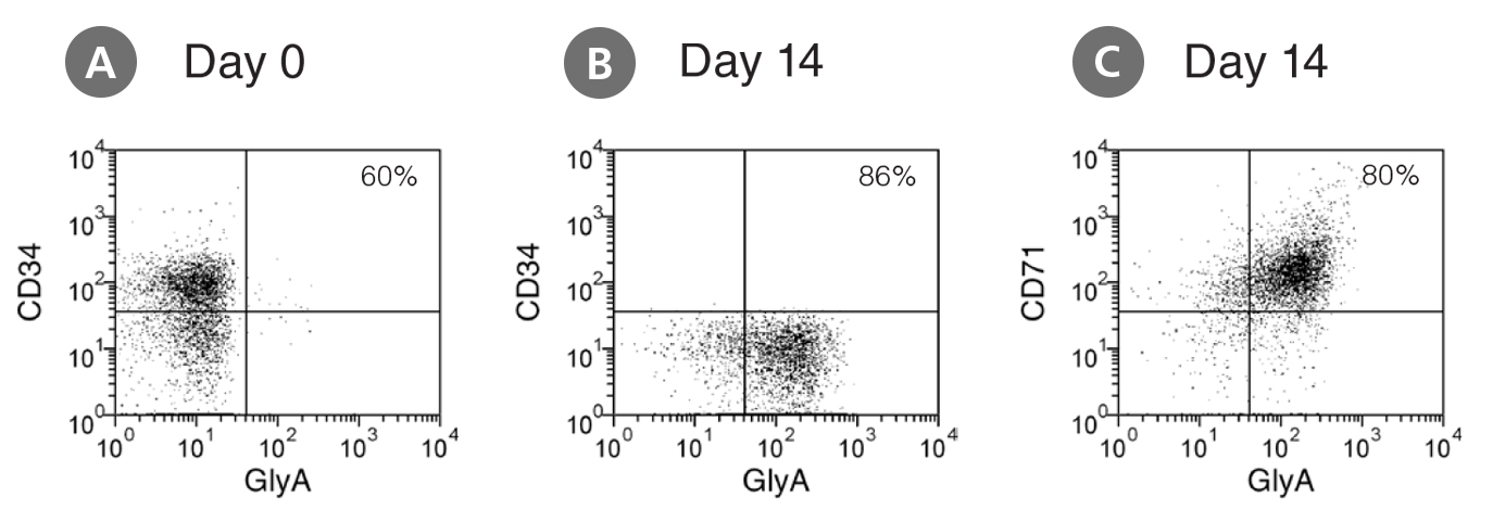
Flow cytometry dot plots showing expression of CD34, CD71, and GlyA before and after culture in StemSpan™

Figure 1. Production of Erythroblasts by Expansion and Lineage Specific Differentiation of CD34+ Human Cord Blood Cells Cultured in StemSpan™ SFEM Containing Erythroid Expansion Supplement

Flow cytometry dot plots showing expression of the HSPC marker CD34 and erythroid markers CD71 and glycophorin-A (GlyA) (A) before culture and (B,C) after 14 days of culture of enriched CD34+ CB cells in StemSpan™ SFEM containing Erythroid Expansion Supplement. The frequency of CD34+ cells declined from ~60% before culture to <0.1% after 14 days. In parallel, erythroid cells gradually accumulated from levels of <1% before culture to >90% by day 14. The bulk of cell population recovered from 14-day culture consisted of CD71+GlyA+ erythroblasts. More immature CD71+GlyA- progenitors and proerythroblasts, as well as more differentiated CD71-/low GlyA+ normoblasts were also present at low frequencies.
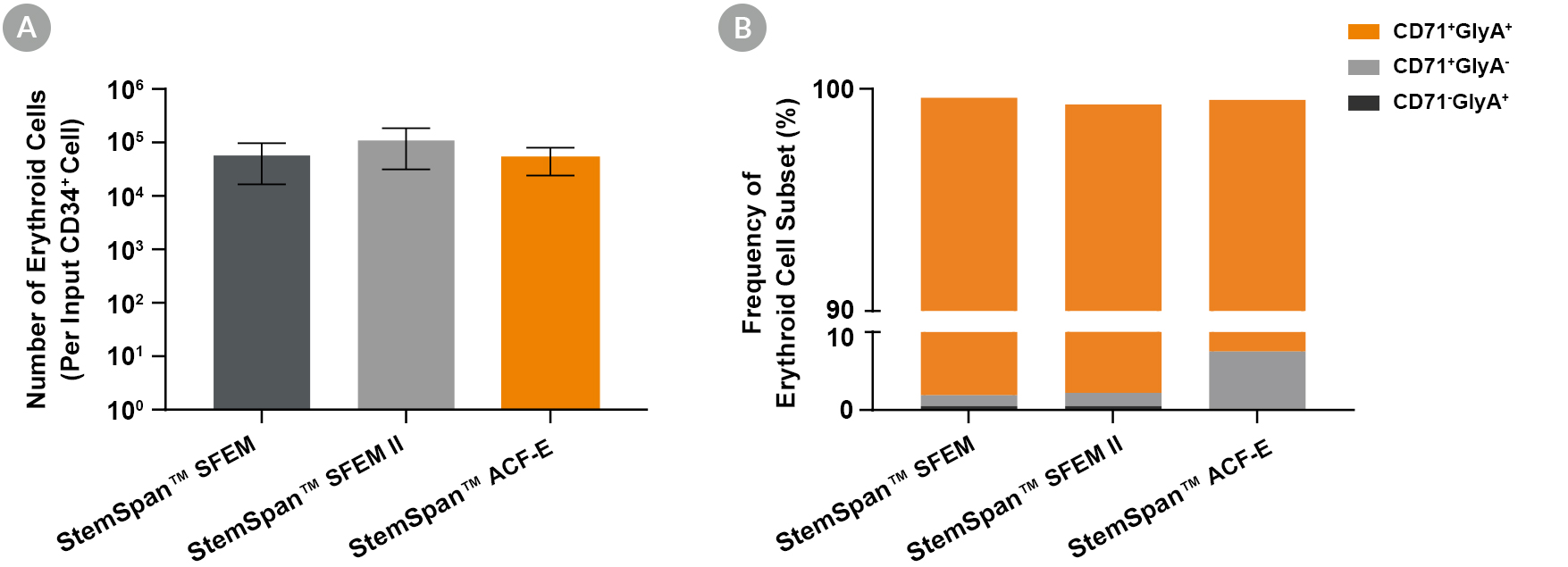
Thousands of Erythroid Cells are Produced Per Input Human CB-Derived CD34+ Cell When Cultured in StemSpan™ Media Containing StemSpan™ Erythroid Expansion Supplement

Figure 2. Production of Erythroid Cells from Human CB-Derived CD34+ Cells Cultured in StemSpan™ Media Containing StemSpan™ Erythroid Expansion Supplement
(A) Average numbers of erythroid cells generated after culturing purified CD34+ CB cells (n = 5) for 14 days in StemSpan™ SFEM (black bars), SFEM II (grey bars), or StemSpan™-ACF Erythroid Expansion Medium (ACF-E, orange bars) media containing StemSpan™ Erythroid Expansion Supplement (Catalog #02692). Shown are the number of erythroid cells that express CD71 and/or Glycophorin A (GlyA) per input CD34+ cell. (B) The percentages of the different erythroid cell subsets generated in these cultures are shown, including CD71+GlyA+ erythroblasts, immature CD71+GlyA- erythroid progenitor cells and pro-erythroblasts, and CD71-/lowGlyA+ normoblasts. All three media supported the generation of thousands of erythroid cells per CB-derived CD34+ cell plated.

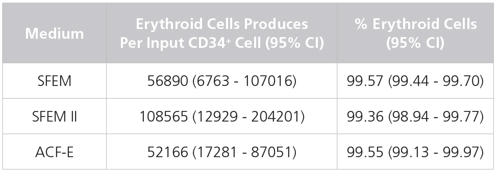
Table 1. Production of Erythroid Cells from Human CB-Derived CD34+ Cells Cultured in StemSpan™ Media Containing StemSpan™ Erythroid Expansion Supplement

Production of Erythroid Cells from Human CB-Derived CD34+ Cells Cultured in StemSpan™ Media Containing StemSpan™ Erythroid Expansion Supplement

Yields and percentages of erythroid cells produced after culturing purified CD34+ CB cells (n = 5) for 14 days in StemSpan™ SFEM, SFEM II or ACF-E media containing Erythroid Expansion Supplement. Erythroid cells were identified by flow cytometry after staining with antibodies against CD71 and GlyA. The % erythroid cells represent the percentage of cells that express CD71 and/or GlyA. *CI: Confidence Interval.

Publications (3)

Stem cell research 2017 Generation of induced pluripotent stem cells (iPSCs) from a hypertrophic cardiomyopathy patient with the pathogenic variant p.Val698Ala in beta-myosin heavy chain (MYH7) gene. S. B. Ross et al.

Abstract

Induced pluripotent stem cells (iPSCs) were generated from peripheral blood mononuclear cells (PBMCs) isolated from the whole blood of a 43-year-old male with hypertrophic cardiomyopathy (HCM) who carries the pathogenic variant p.Val698Ala in beta-myosin heavy chain (MYH7). Patient-derived PBMCs were reprogrammed using non-integrative episomal vectors containing reprogramming factors OCT4, SOX2, LIN28, KLF4 and L-MYC. iPSCs were shown to express pluripotent markers, have trilineage differentiation potential, carry the pathogenic MYH7 variant p.Val698Ala, have a normal karyotype and no longer carry the episomal reprogramming vector. This line is useful for studying the link between variants in MYH7 and the pathogenesis of HCM.
Stem cell research 2017 Peripheral blood derived induced pluripotent stem cells (iPSCs) from a female with familial hypertrophic cardiomyopathy. S. B. Ross et al.

Abstract

Induced pluripotent stem cells (iPSCs) were generated from peripheral blood mononuclear cells (PBMCs) obtained from a 62-year-old female with familial hypertrophic cardiomyopathy (HCM). PBMCs were reprogrammed to a pluripotent state following transfection with non-integrative episomal vectors carrying reprogramming factors OCT4, SOX2, LIN28, KLF4 and L-MYC. iPSCs were shown to express pluripotency markers, possess trilineage differentiation potential, carry rare variants identified in DNA isolated directly from the patient's whole blood, have a normal karyotype and no longer carry episomal vectors for reprogramming. This line is a useful resource for identifying unknown genetic causes of HCM.
Science translational medicine 2016 OCT Selection-free genome editing of the sickle mutation in human adult hematopoietic stem/progenitor cells. M. A. DeWitt et al.

Abstract

Genetic diseases of blood cells are prime candidates for treatment through ex vivo gene editing of CD34+ hematopoietic stem/progenitor cells (HSPCs), and a variety of technologies have been proposed to treat these disorders. Sickle cell disease (SCD) is a recessive genetic disorder caused by a single-nucleotide polymorphism in the $\beta$-globin gene (HBB). Sickle hemoglobin damages erythrocytes, causing vasoocclusion, severe pain, progressive organ damage, and premature death. We optimize design and delivery parameters of a ribonucleoprotein (RNP) complex comprising Cas9 protein and unmodified single guide RNA, together with a single-stranded DNA oligonucleotide donor (ssODN), to enable efficient replacement of the SCD mutation in human HSPCs. Corrected HSPCs from SCD patients produced less sickle hemoglobin RNA and protein and correspondingly increased wild-type hemoglobin when differentiated into erythroblasts. When engrafted into immunocompromised mice, ex vivo treated human HSPCs maintain SCD gene edits throughout 16 weeks at a level likely to have clinical benefit. These results demonstrate that an accessible approach combining Cas9 RNP with an ssODN can mediate efficient HSPC genome editing, enables investigator-led exploration of gene editing reagents in primary hematopoietic stem cells, and suggests a path toward the development of new gene editing treatments for SCD and other hematopoietic diseases.
View All Publications
Top