
STEMdiff™ Pancreatic Progenitor Kit


STEMdiff™ Pancreatic Progenitor Kit has been optimized for differentiation of cells maintained in mTeSR™1 (Catalog #85850).
• Efficient and reproducible differentiation of multiple hPSC lines to pancreatic progenitor cells expressing PDX-1, NKX6.1, and SOX9
• Serum-free medium
• Compatible with multiple human ES and iPS cell lines maintained in mTeSR™1
- STEMdiff™ Endoderm Basal Medium, 100 mL
- STEMdiff™ Pancreatic Stage 2 - 4 Basal Medium, 265 mL
- STEMdiff™ Definitive Endoderm Supplement MR (100X), 350 µL
- STEMdiff™ Definitive Endoderm Supplement CJ (100X), 1.1 mL
- STEMdiff™ Pancreatic Supplement 2A, 240 µL
- STEMdiff™ Pancreatic Supplement 2B, 720 µL
- STEMdiff™ Pancreatic Supplement 3, 720 µL
- STEMdiff™ Pancreatic Supplement 4, 1.2 mL
| Document Type | 产品名称 | Catalog # | Lot # | 语言 |
|---|---|---|---|---|
| Product Information Sheet 1 | STEMdiff™ Pancreatic Progenitor Kit | 05120 | Component #05113: Lot 18D89658 and higher | English |
| Product Information Sheet 2 | STEMdiff™ Pancreatic Progenitor Kit | 05120 | Component #05113: Lot 18D89658 and higher | English |
| Safety Data Sheet 1 | STEMdiff™ Pancreatic Progenitor Kit | 05120 | All | English |
| Safety Data Sheet 2 | STEMdiff™ Pancreatic Progenitor Kit | 05120 | All | English |
| Safety Data Sheet 3 | STEMdiff™ Pancreatic Progenitor Kit | 05120 | All | English |
| Safety Data Sheet 4 | STEMdiff™ Pancreatic Progenitor Kit | 05120 | All | English |
| Safety Data Sheet 5 | STEMdiff™ Pancreatic Progenitor Kit | 05120 | All | English |
| Safety Data Sheet 6 | STEMdiff™ Pancreatic Progenitor Kit | 05120 | All | English |
| Safety Data Sheet 7 | STEMdiff™ Pancreatic Progenitor Kit | 05120 | All | English |
Data
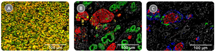
Figure 1. Pancreatic Progenitor Cells can Mature into Endocrine and Exocrine Cells
A) Representative image showing pancreatic progenitor cells expressing PDX-1 (red) and NKX6.1 (green). Yellow staining indicates co‑expression of both markers in the majority of cells as is observed in the developing human pancreas.1 (B, C) Cells transplanted into mice can mature into endocrine and exocrine cells.
B) shows endocrine clusters expressing synaptophysin (red) surrounded by ductal structures expressing CK-19 (green).
C) Shows islet-like structures containing monohormonal cells that individually express insulin (red), glucagon (green) or somatostatin (blue). Data in (B, C) are from the laboratory of Dr. Timothy J. Kieffer (University of British Columbia, Vancouver, Canada).
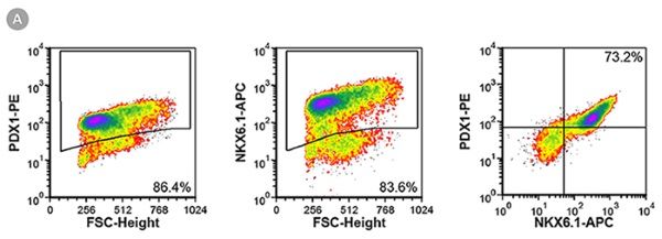
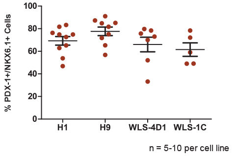
Figure 2. The STEMdiff™ Pancreatic Progenitor Kit Functions Efficiently Across Multiple hPSC Lines
PDX-1 and NKX6.1 expression measured in pancreatic progenitor cells derived from four different hPSC lines (H1, H9, WLS-4D1 and WLS-1C) at the end of Stage 4. (A) Representative flow cytometry plots show PDX-1 and NKX6.1 co-expression in differentiated H9 cells. (B) Quantitative data for PDX-1/NKX6.1 co-expression in two human ES (H1 and H9) and two human iPS (WLS-4D1 and WLS-1C) cell lines (n = 5-10 per cell line). Data are plotted as individual points representing the mean of duplicates within a single experiment. The horizontal line represents the mean of all experiments, with error bars indicating the standard error of the mean (SEM). The average efficiency of pancreatic progenitor differentiation ranges from 61.5% to 77.7% depending on the cell line.
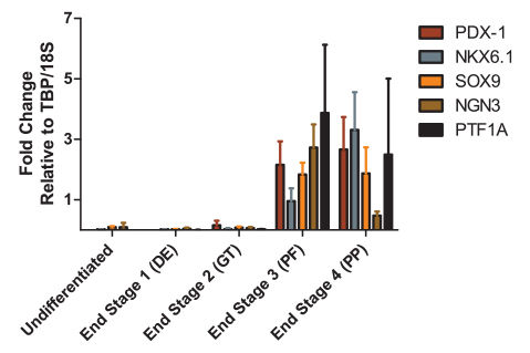
Figure 3. Gene Expression Profile is Indicative of Transition to Pancreatic Progenitor Cells
Gene expression profile at the end of each stage of differentiation for key markers of pancreatic progenitor cells. Expression was normalized to 18S ribosomal RNA and TATA Binding Protein (TBP). Data are the mean ± SEM for 3 - 5 experiments. Expression pattern is consistent with published data.²
1. Riedel M et al. (2012) Immunohistochemical characterization of cells co-producing insulin and glucagon in the developing human pancreas. Diabetologia 55(2): 372-81.
2. Rezania A et al. (2014) Reversal of diabetes with insulin-producing cells derived in vitro from human pluripotent stem cells. Nat Biotechnol 32(11): 1121-33.
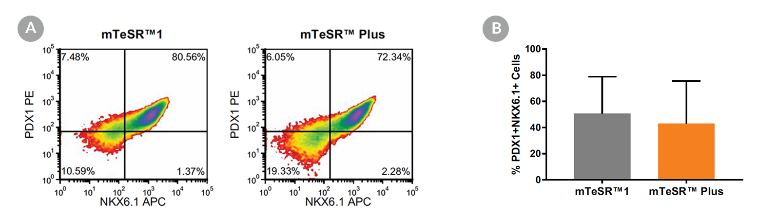
Figure 4. Generation of Pancreatic Progenitors from hPSCs Maintained in mTeSR™ Plus
(A) Representative density plots showing PDX-1 and NKX6.1 expression in cells cultured in mTeSR™1 (daily feeds) or mTeSR™ Plus (restricted feeds), following differentiation using the STEMdiff™ Pancreatic Progenitor Kit. (B) Quantitative analysis of pancreatic progenitor formation in multiple hPS (H9, STiPS-M001, WLS-1C) cell lines maintained with mTeSR™1 or mTeSR™ Plus as measured by co-expression of PDX-1 and NKX6.1. Data are expressed as the mean percentage of cells (± SEM) expressing both markers; n=3.