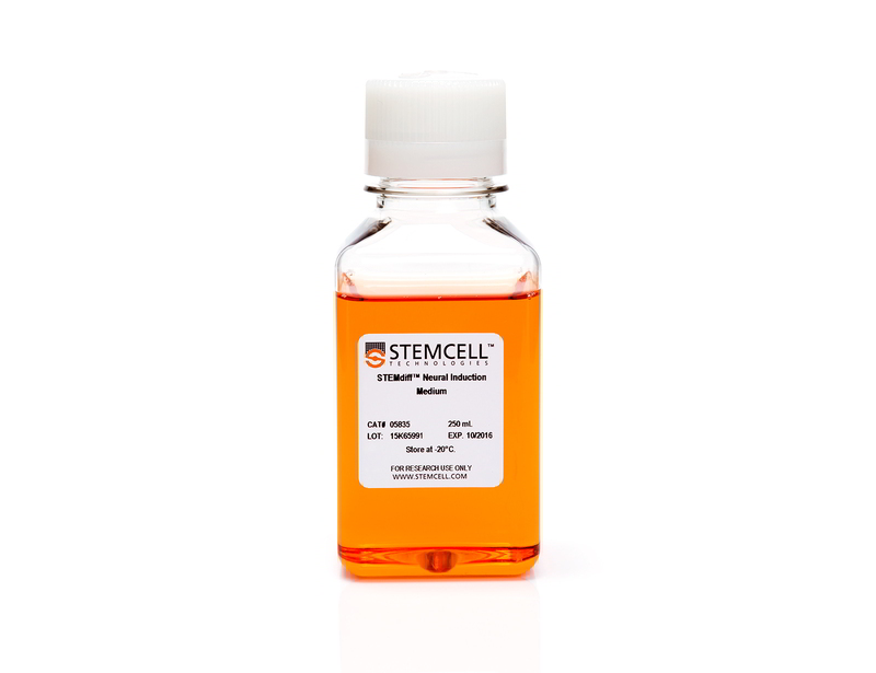
STEMdiff™ Neural Induction Medium
Defined, serum-free medium for neural induction of human ES and iPS cells
概要
STEMdiff™ Neural Induction Medium is a defined, serum-free medium for the neural induction of human embryonic stem (ES) cells and induced pluripotent stem (iPS) cells. This medium enables highly efficient generation of neural progenitor cell using either embryoid body- or monolayer culture-based protocols.
Advantages
• Defined and serum-free
• Rapid and efficient neural induction
• Compatible with both embryoid body and monolayer culture protocols
• Reproducible differentiation of multiple ES cell and iPS cell lines maintained in mTeSR™ 1
• Convenient, user-friendly format and protocols
• Rapid and efficient neural induction
• Compatible with both embryoid body and monolayer culture protocols
• Reproducible differentiation of multiple ES cell and iPS cell lines maintained in mTeSR™ 1
• Convenient, user-friendly format and protocols
Components
- STEMdiff™ Neural Induction Medium (Catalog #05835)
- STEMdiff™ Neural Induction Medium, 2 x 250 mL (Catalog #05839)
Subtype
Specialized Media
Cell Type
Neural Cells, PSC-Derived, Neural Stem and Progenitor Cells, Pluripotent Stem Cells
Species
Human
Application
Cell Culture, Differentiation
Brand
STEMdiff
Area of Interest
Disease Modeling, Drug Discovery and Toxicity Testing, Neuroscience, Stem Cell Biology
Formulation
Serum-Free
技术资料
| Document Type | 产品名称 | Catalog # | Lot # | 语言 |
|---|---|---|---|---|
| Product Information Sheet | STEMdiff™ Neural Induction Medium | 05835, 05839 | All | English |
| Manual | STEMdiff™ Neural Induction Medium | 05835 | All | English |
| Safety Data Sheet | STEMdiff™ Neural Induction Medium | 05835, 05839 | All | English |
数据及文献
Publications (38)
Biosensors 2019 apr
A Novel Toolkit for Characterizing the Mechanical and Electrical Properties of Engineered Neural Tissues.
Abstract
Abstract
We have designed and validated a set of robust and non-toxic protocols for directly evaluating the properties of engineered neural tissue. These protocols characterize the mechanical properties of engineered neural tissues and measure their electrophysical activity. The protocols obtain elastic moduli of very soft fibrin hydrogel scaffolds and voltage readings from motor neuron cultures. Neurons require soft substrates to differentiate and mature, however measuring the elastic moduli of soft substrates remains difficult to accurately measure using standard protocols such as atomic force microscopy or shear rheology. Here we validate a direct method for acquiring elastic modulus of fibrin using a modified Hertz model for thin films. In this method, spherical indenters are positioned on top of the fibrin samples, generating an indentation depth that is then correlated with elastic modulus. Neurons function by transmitting electrical signals to one another and being able to assess the development of electrical signaling serves is an important verification step when engineering neural tissues. We then validated a protocol wherein the electrical activity of motor neural cultures is measured directly by a voltage sensitive dye and a microplate reader without causing damage to the cells. These protocols provide a non-destructive method for characterizing the mechanical and electrical properties of living spinal cord tissues using novel biosensing methods.
Human molecular genetics 2019
Multiplication of the SNCA locus exacerbates neuronal nuclear aging.
Abstract
Abstract
Human-induced Pluripotent Stem Cell (hiPSC)-derived models have advanced the study of neurodegenerative diseases, including Parkinson's disease (PD). While age is the strongest risk factor for these disorders, hiPSC-derived models represent rejuvenated neurons. We developed hiPSC-derived Aged dopaminergic and cholinergic neurons to model PD and related synucleinopathies. Our new method induces aging through a `semi-natural' process, by passaging multiple times at the Neural Precursor Cell stage, prior to final differentiation. Characterization of isogenic hiPSC-derived neurons using heterochromatin and nuclear envelope markers, as well as DNA damage and global DNA methylation, validated our age-inducing method. Next, we compared neurons derived from a patient with SNCA-triplication (SNCA-Tri) and a Control. The SNCA-Tri neurons displayed exacerbated nuclear aging, showing advanced aging signatures already at the Juvenile stage. Noteworthy, the Aged SNCA-Tri neurons showed more $\alpha$-synuclein aggregates per cell versus the Juvenile. We suggest a link between the effects of aging and SNCA overexpression on neuronal nuclear architecture.
Translational psychiatry 2019
Comparative characterization of human induced pluripotent stem cells (hiPSC) derived from patients with schizophrenia and autism.
Abstract
Abstract
Human induced pluripotent stem cells (hiPSC) provide an attractive tool to study disease mechanisms of neurodevelopmental disorders such as schizophrenia. A pertinent problem is the development of hiPSC-based assays to discriminate schizophrenia (SZ) from autism spectrum disorder (ASD) models. Healthy control individuals as well as patients with SZ and ASD were examined by a panel of diagnostic tests. Subsequently, skin biopsies were taken for the generation, differentiation, and testing of hiPSC-derived neurons from all individuals. SZ and ASD neurons share a reduced capacity for cortical differentiation as shown by quantitative analysis of the synaptic marker PSD95 and neurite outgrowth. By contrast, pattern analysis of calcium signals turned out to discriminate among healthy control, schizophrenia, and autism samples. Schizophrenia neurons displayed decreased peak frequency accompanied by increased peak areas, while autism neurons showed a slight decrease in peak amplitudes. For further analysis of the schizophrenia phenotype, transcriptome analyses revealed a clear discrimination among schizophrenia, autism, and healthy controls based on differentially expressed genes. However, considerable differences were still evident among schizophrenia patients under inspection. For one individual with schizophrenia, expression analysis revealed deregulation of genes associated with the major histocompatibility complex class II (MHC class II) presentation pathway. Interestingly, antipsychotic treatment of healthy control neurons also increased MHC class II expression. In conclusion, transcriptome analysis combined with pattern analysis of calcium signals appeared as a tool to discriminate between SZ and ASD phenotypes in vitro.
Cell stem cell 2017 MAY
Nicotinamide Ameliorates Disease Phenotypes in a Human iPSC Model of Age-Related Macular Degeneration.
Abstract
Abstract
Age-related macular degeneration (AMD) affects the retinal pigment epithelium (RPE), a cell monolayer essential for photoreceptor survival, and is the leading cause of vision loss in the elderly. There are no disease-altering therapies for dry AMD, which is characterized by accumulation of subretinal drusen deposits and complement-driven inflammation. We report the derivation of human-induced pluripotent stem cells (hiPSCs) from patients with diagnosed AMD, including two donors with the rare ARMS2/HTRA1 homozygous genotype. The hiPSC-derived RPE cells produce several AMD/drusen-related proteins, and those from the AMD donors show significantly increased complement and inflammatory factors, which are most exaggerated in the ARMS2/HTRA1 lines. Using a panel of AMD biomarkers and candidate drug screening, combined with transcriptome analysis, we discover that nicotinamide (NAM) ameliorated disease-related phenotypes by inhibiting drusen proteins and inflammatory and complement factors while upregulating nucleosome, ribosome, and chromatin-modifying genes. Thus, targeting NAM-regulated pathways is a promising avenue for developing therapeutics to combat AMD.
Development (Cambridge, England) 2017 MAR
A process engineering approach to increase organoid yield.
Abstract
Abstract
Temporal manipulation of the in vitro environment and growth factors can direct differentiation of human pluripotent stem cells into organoids - aggregates with multiple tissue-specific cell types and three-dimensional structure mimicking native organs. A mechanistic understanding of early organoid formation is essential for improving the robustness of these methods, which is necessary prior to use in drug development and regenerative medicine. We investigated intestinal organoid emergence, focusing on measurable parameters of hindgut spheroids, the intermediate step between definitive endoderm and mature organoids. We found that 13{\%} of spheroids were pre-organoids that matured into intestinal organoids. Spheroids varied by several structural parameters: cell number, diameter and morphology. Hypothesizing that diameter and the morphological feature of an inner mass were key parameters for spheroid maturation, we sorted spheroids using an automated micropipette aspiration and release system and monitored the cultures for organoid formation. We discovered that populations of spheroids with a diameter greater than 75 $\mu$m and an inner mass are enriched 1.5- and 3.8-fold for pre-organoids, respectively, thus providing rational guidelines towards establishing a robust protocol for high quality intestinal organoids.
Stem Cell Reports 2017 MAR
Reversal of Phenotypic Abnormalities by CRISPR/Cas9-Mediated Gene Correction in Huntington Disease Patient-Derived Induced Pluripotent Stem Cells
Abstract
Abstract
Huntington disease (HD) is a dominant neurodegenerative disorder caused by a CAG repeat expansion in HTT. Here we report correction of HD human induced pluripotent stem cells (hiPSCs) using a CRISPR-Cas9 and piggyBac transposon-based approach. We show that both HD and corrected isogenic hiPSCs can be differentiated into excitable, synaptically active forebrain neurons. We further demonstrate that phenotypic abnormalities in HD hiPSC-derived neural cells, including impaired neural rosette formation, increased susceptibility to growth factor withdrawal, and deficits in mitochondrial respiration, are rescued in isogenic controls. Importantly, using genome-wide expression analysis, we show that a number of apparent gene expression differences detected between HD and non-related healthy control lines are absent between HD and corrected lines, suggesting that these differences are likely related to genetic background rather than HD-specific effects. Our study demonstrates correction of HD hiPSCs and associated phenotypic abnormalities, and the importance of isogenic controls for disease modeling using hiPSCs.


