STEMdiff™ Mesenchymal Progenitor Kit
Defined culture kit for derivation and expansion of mesenchymal progenitor cells
概要
STEMdiff™ Mesenchymal Progenitor Kit is a defined culture kit consisting of animal component-free (ACF) induction medium, expansion medium, and attachment substrate. It is optimized for the derivation of cells with mesenchymal progenitor cell (MPC)-like properties from human embryonic stem (ES) cells or induced pluripotent stem (iPS) cells. This kit provides a complete workflow of defined reagents for derivation and expansion of human ES- or iPS-derived MPCs.
• Serum- and animal component-free formulation
• Efficient and reproducible generation of MPCs from human ES and iPS cell lines
• Rapid derivation of MPCs in 3 weeks
• Generates MPCs capable of long-term expansion and differentiation to adipocytes, osteoblasts, and chondrocytes
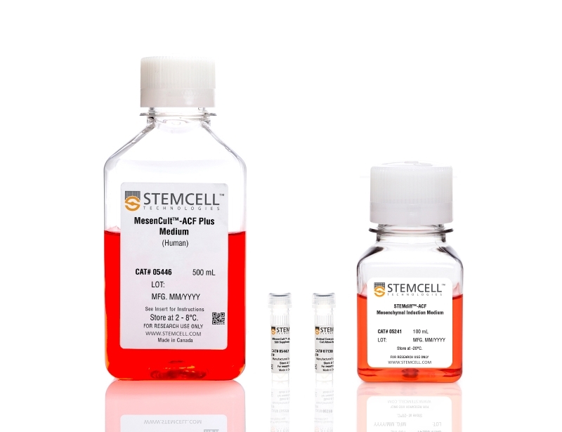
- STEMdiff™-ACF Mesenchymal Induction Medium, 100 mL
- MesenCult™-ACF Plus Medium, 500 mL
- MesenCult™-ACF Plus 500X Supplement, 1 mL
- Animal Component-Free Cell Attachment Substrate, 1 mL
Mesenchymal Cells, PSC-Derived
Cell Culture, Differentiation
数据及文献
Publications (6)
Biochemical and biophysical research communications 2020 may
Induced pluripotency and spontaneous reversal of cellular aging in supercentenarian donor cells.
J. Lee et al.
Abstract
Supercentenarians (≥110-year-old, SC) are a uniquely informative population not only because they surpass centenarians in age, but because they appear to age more slowly with fewer incidences of chronic age-related disease than centenarians. We reprogramed donor B-lymphoblastoid cell lines (LCL) derived from a 114-year-old (SC), a 43-year-old healthy disease-free control (HDC) and an 8-year-old with a rapid aging disease (Hutchinson-Gilford progeria syndrome (HGPS)) and compared SC-iPSC to HDC-iPSC and HGPS-iPSCs. Reprogramming to pluripotency was confirmed by pluripotency marker expression and differentiation to 3 germ-layers. Each iPSC clone differentiated efficiently to mesenchymal progenitor cells (MPC) as determined by surface marker expression and RNAseq analysis. We identified supercentenarian and HGPS associated gene expression patterns in the differentiated MPC lines that were not evident in the parental iPSC lines. Importantly, telomere length resetting occurred in iPSC from all donors albeit at a lower incidence in supercentenarian iPSCs. These data indicate the potential to use reprogramming to reset both developmental state and cellular age in the oldest of the old." We anticipate that supercentenarian iPSC and their differentiated derivatives will be valuable tools for studying the underlying mechanisms of extreme longevity and disease resistance."
Journal of cellular and molecular medicine 2020 mar
Intravenous administration of iPS-MSCSPIONs mobilized into CKD parenchyma and effectively preserved residual renal function in CKD rat.
J.-J. Sheu et al.
Abstract
This study traced intravenously administered induced pluripotent stem cell (iPSC)-derived mesenchymal stem cells (MSC) and assessed the impact of iPSC-MSC on preserving renal function in SD rat after 5/6 nephrectomy. The results of in vitro study showed that FeraTrack™Direct contrast particles (ie intracellular magnetic labelling) in the iPSC-MSC (ie iPS-MSCSPIONs ) were clearly identified by Prussian blue stain. Adult-male SD rats (n = 40) were categorized into group 1 (SC), group 2 [SC + iPS-MSCSPIONs (1.0 × 10⁶ cells)/intravenous administration post-day-14 CKD procedure], group 3 (CKD), group 4 [CKD + iPS-MSCSPIONs (0.5 × 10⁶ cells)] and group 5 [CKD + iPS-MSCSPIONs (1.0 × 10⁶ cells)]. By day-15 after CKD induction, abdominal MRI demonstrated that iPS-MSCSPIONs were only in the CKD parenchyma of groups 4 and 5. By day 60, the creatinine level/ratio of urine protein to urine creatinine/kidney injury score (by haematoxylin and eosin stain)/fibrotic area (Masson's trichrome stain)/IF microscopic finding of kidney injury molecule-1 expression was lowest in groups 1 and 2, highest in group 3, and significantly higher in group 4 than in group 5, whereas IF microscopic findings of podocyte components (ZO-1/synaptopodin) and protein levels of anti-apoptosis ((Bad/Bcl-xL/Bcl-2) exhibited an opposite pattern to creatinine level among the five groups (all P {\textless} .0001). The protein expressions of cell-proliferation signals (PI3K/p-Akt/m-TOR, p-ERK1/2, FOXO1/GSK3$\beta$/p90RSK), apoptotic/DNA-damage (Bax/caspases8-10/cytosolic-mitochondria) and inflammatory (TNF-$\alpha$/TNFR1/TRAF2/NF-$\kappa$B) biomarkers displayed an identical pattern to creatinine level among the five groups (all P {\textless} .0001). The iPS-MSCSPIONs that were identified only in CKD parenchyma effectively protected the kidney against CKD injury.
Stem cells translational medicine 2020 jul
Precision installation of a highly efficient suicide gene safety switch in human induced pluripotent stem cells.
Z.-D. Shi et al.
Abstract
Human pluripotent stem cells including induced pluripotent stem cells (iPSCs) and embryonic stem cells hold great promise for cell-based therapies, but safety concerns that complicate consideration for routine clinical use remain. Installing a safety switch" based on the inducible caspase-9 (iCASP9) suicide gene system should offer added control over undesirable cell replication or activity. Previous studies utilized lentiviral vectors to integrate the iCASP9 system into T cells and iPSCs. This method results in random genomic insertion of the suicide switch and inefficient killing of the cells after the switch is "turned on" with a small molecule (eg AP1903). To improve the safety and efficiency of the iCASP9 system for use in iPSC-based therapy we precisely installed the system into a genomic safe harbor the AAVS1 locus in the PPP1R12C gene. We then evaluated the efficiencies of different promoters to drive iCASP9 expression in human iPSCs. We report that the commonly used EF1$\alpha$ promoter is silenced in iPSCs and that the endogenous promoter of the PPP1R12C gene is not strong enough to drive high levels of iCASP9 expression. However the CAG promoter induces strong and stable iCASP9 expression in iPSCs and activation of this system with AP1903 leads to rapid killing and complete elimination of iPSCs and their derivatives including MSCs and chondrocytes in vitro. Furthermore iPSC-derived teratomas shrank dramatically or were completely eliminated after administration of AP1903 in mice. Our data suggest significant improvements on existing iCASP9 suicide switch technologies and may serve as a guide to other groups seeking to improve the safety of stem cell-based therapies."
BMC cancer 2019 jun
miR-520d-5p can reduce the mutations in hepatoma cancer cells and iPSCs-derivatives.
N. Miura et al.
Abstract
BACKGROUND Human microRNAs (miRNAs) have diverse functions in biology, and play a role in nearly every biological process. Here we report that miR-520d-5p (520d-5p) causes undifferentiated cancer cells to adopt benign or normal status in vivo in immunodeficient mice via demethylation and P53 upregulation. Further we found that 520-5p causes normal cells to elongate cellular lifetime and mesenchymal stem cell-like status with CD105 positivity. We hypothesized that ectopic 520d-5p expression reduced mutations in undifferentiated type of hepatoma (HLF) cells through synergistic modulation of methylation-related enzymatic expression. METHODS To examine whether there were any changes in mutation status in cells treated with 520d-5p, we performed next generation sequencing (NGS) in HLF cells and human iPSC-derivative cells in pre-mesenchymal stem cell status. We analyzed the data using both genome-wide and individual gene function approaches. RESULTS 520d-5p induced a shift towards a wild type or non-malignant phenotype, which was regulated by nucleotide mutations in both HLF cells and iPSCs. Further, 520d-5p reduced mutation levels in both the whole genome and genomic fragment assemblies. CONCLUSIONS Cancer cell genomic mutations cannot be repaired in most contexts. However, these findings suggest that applied development of 520d-5p would allow new approaches to cancer research and improve the quality of iPSCs used in regenerative medicine.
Nature 2018
Linking a cell-division gene and a suicide gene to define and improve cell therapy safety.
Q. Liang et al.
Abstract
Human pluripotent cell lines hold enormous promise for the development of cell-based therapies. Safety, however, is a crucial prerequisite condition for clinical applications. Numerous groups have attempted to eliminate potentially harmful cells through the use of suicide genes1, but none has quantitatively defined the safety level of transplant therapies. Here, using genome-engineering strategies, we demonstrate the protection of a suicide system from inactivation in dividing cells. We created a transcriptional link between the suicide gene herpes simplex virus thymidine kinase (HSV-TK) and a cell-division gene (CDK1); this combination is designated the safe-cell system. Furthermore, we used a mathematical model to quantify the safety level of the cell therapy as a function of the number of cells that is needed for the therapy and the type of genome editing that is performed. Even with the highly conservative estimates described here, we anticipate that our solution will rapidly accelerate the entry of cell-based medicine into the clinic.
Stem cells translational medicine 2018
Ex Vivo Expansion of CD34+ CD90+ CD49f+ Hematopoietic Stem and Progenitor Cells from Non-Enriched Umbilical Cord Blood with Azole Compounds.
S. Bari et al.
Abstract
Umbilical cord blood (UCB) transplants in adults have slower hematopoietic recovery compared to bone marrow (BM) or peripheral blood (PB) stem cells mainly due to low number of total nucleated cells and hematopoietic stem and progenitor cells (HSPC). As such in this study, we aimed to perform ex vivo expansion of UCB HSPC from non-enriched mononucleated cells (MNC) using novel azole-based small molecules. Freshly-thawed UCB-MNC were cultured in expansion medium supplemented with small molecules and basal cytokine cocktail. The effects of the expansion protocol were measured based on in vitro and in vivo assays. The proprietary library of {\textgreater}50 small molecules were developed using structure-activity-relationship studies of SB203580, a known p38-MAPK inhibitor. A particular analog, C7, resulted in 1,554.1 ± 27.8-fold increase of absolute viable CD45+ CD34+ CD38- CD45RA- progenitors which was at least 3.7-fold higher than control cultures (p {\textless} .001). In depth phenotypic analysis revealed {\textgreater}600-fold expansion of CD34+ /CD90+ /CD49f+ rare HSPCs coupled with significant (p {\textless} .01) increase of functional colonies from C7 treated cells. Transplantation of C7 expanded UCB grafts to immunodeficient mice resulted in significantly (p {\textless} .001) higher engraftment of human CD45+ and CD45+ CD34+ cells in the PB and BM by day 21 compared to non-expanded and cytokine expanded grafts. The C7 expanded grafts maintained long-term human multilineage chimerism in the BM of primary recipients with sustained human CD45 cell engraftment in secondary recipients. In conclusion, a small molecule, C7, could allow for clinical development of expanded UCB grafts without pre-culture stem cell enrichment that maintains in vitro and in vivo functionality. Stem Cells Translational Medicine 2018;7:376-393.
View All Publications