STEMdiff™ Kidney Organoid Kit
Kidney organoids generated with STEMdiff™ Kidney Organoid Kit are tested for compatibility with phenotypic high-throughput assays such as nephrotoxic compound screening. They also provide a relevant and convenient model for studies related to developmental biology and disease modeling.
⦁ SIMPLE. Minimizes culture manipulations with a two-stage culture system and easy-to-follow protocol.
⦁ RELIABLE. Provides low experimental variability through an optimized formulation and rigorous quality controls.
⦁ HIGH THROUGHPUT. Reproducibly generates organoids in 96- and 384-well formats.
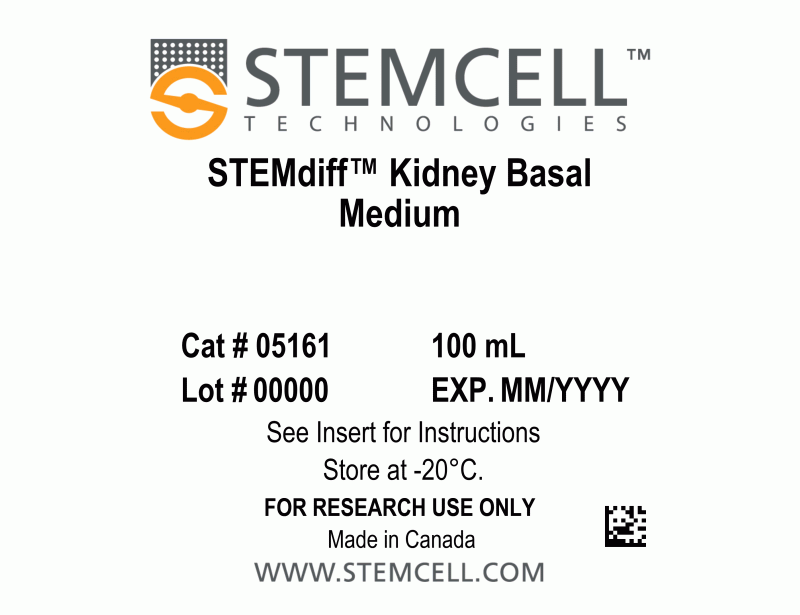
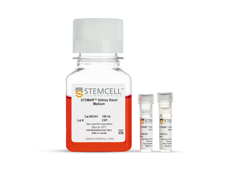
- STEMdiff™ Kidney Basal Medium, 100 mL
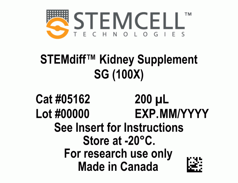
- STEMdiff™ Kidney Supplement SG (100X), 200 µL
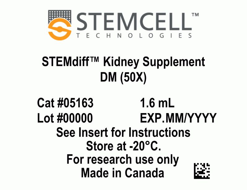
- STEMdiff™ Kidney Supplement DM (50X), 1.6 mL
| Document Type | 产品名称 | Catalog # | Lot # | 语言 |
|---|---|---|---|---|
| Product Information Sheet | STEMdiff™ Kidney Organoid Kit | 05160 | All | English |
| Manual | STEMdiff™ Kidney Organoid Kit | 05160 | All | English |
| Safety Data Sheet 1 | STEMdiff™ Kidney Organoid Kit | 05160 | All | English |
| Safety Data Sheet 2 | STEMdiff™ Kidney Organoid Kit | 05160 | All | English |
| Safety Data Sheet 3 | STEMdiff™ Kidney Organoid Kit | 05160 | All | English |
Data
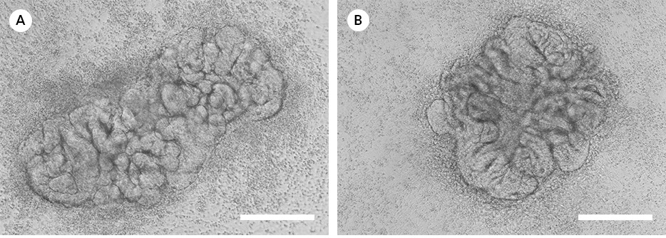
Figure 1. Representative Images of Kidney Organoids
The STEMdiff™ Kidney Organoid Kit facilitates the directed differentiation of hPSCs to form kidney organoids that model the developing nephron. Pictured are human kidney organoids grown using the STEMdiff™ Kidney Organoid Kit differentiated from (A) iPS (WLS-1C) or (B) ES (H9) cells and imaged on day 12 and day 18 of differentiation, respectively.
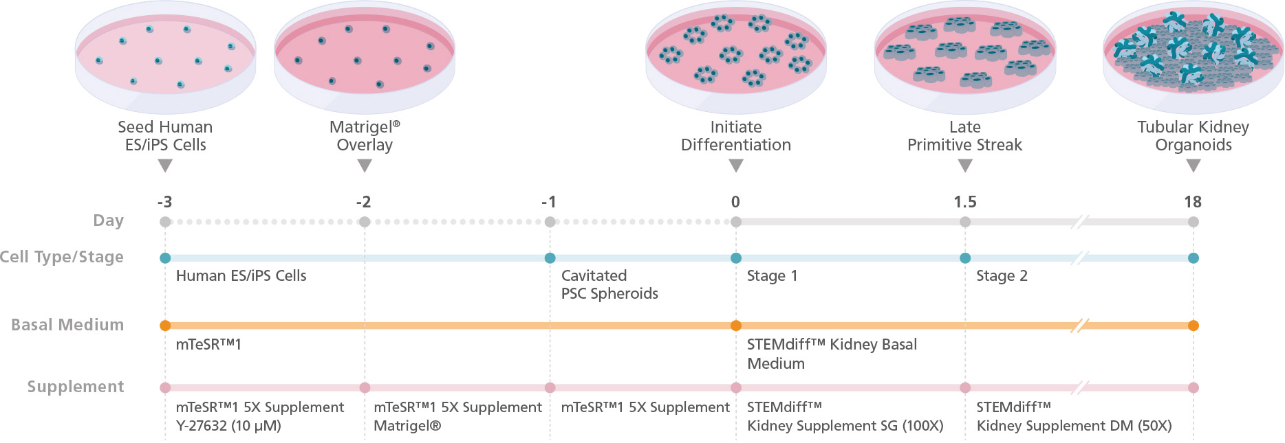
Figure 2. Schematic for Differentiation from hPSCs to Human Kidney Organoids with the STEMdiff™ Kidney Organoid Kit
hPSC cultures progress through a simple two-stage process to generate kidney organoids. hPSCs are first plated and overlaid with Corning® Matrigel® to form cavitated spheroids. On the following day (Day 0), differentiation of cavitated PSC spheroids is initiated by changing medium from mTeSR™1 to STEMdiff™ Kidney Organoid Kit. During the next 18 days, the cells are directed through stages of the late primitive streak, intermediate mesoderm, and metanephric mesoderm to give rise to kidney organoids that are composed of podocytes, proximal and distal tubules, and an associated endothelium and mesenchyme.
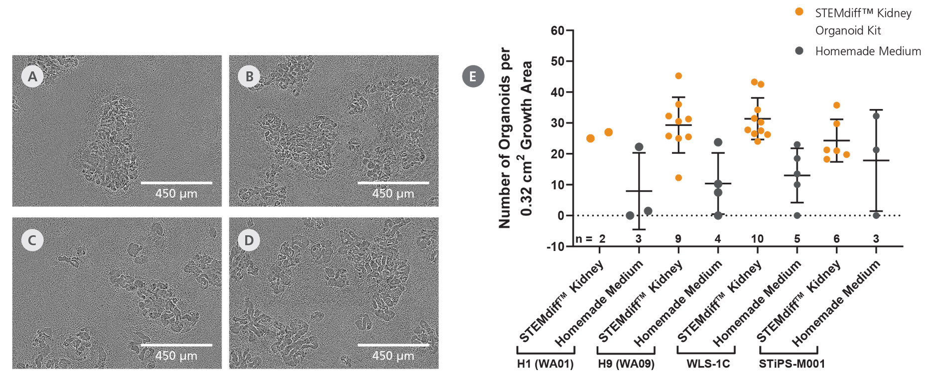
Figure 3. Efficient Differentiation of Human Pluripotent Stem Cells into Self-Organizing Kidney Organoids
The STEMdiff™ Kidney Organoid Kit enables high-efficiency generation of kidney organoids from both ES (A, H1; B, H9) and iPS (C, WLS-1C; D, STiPS-M001) cell lines. (E) Kidney organoids were grown using the STEMdiff™ Kidney Organoid Kit or using home-made medium, and the average number of organoids per well of a 96-well plate was quantified on Day 18. All four tested cell lines were capable of differentiating into self-organizing kidney organoids that form convoluted tubular structures with high efficiency (mean ± SD, n ≥ 2).
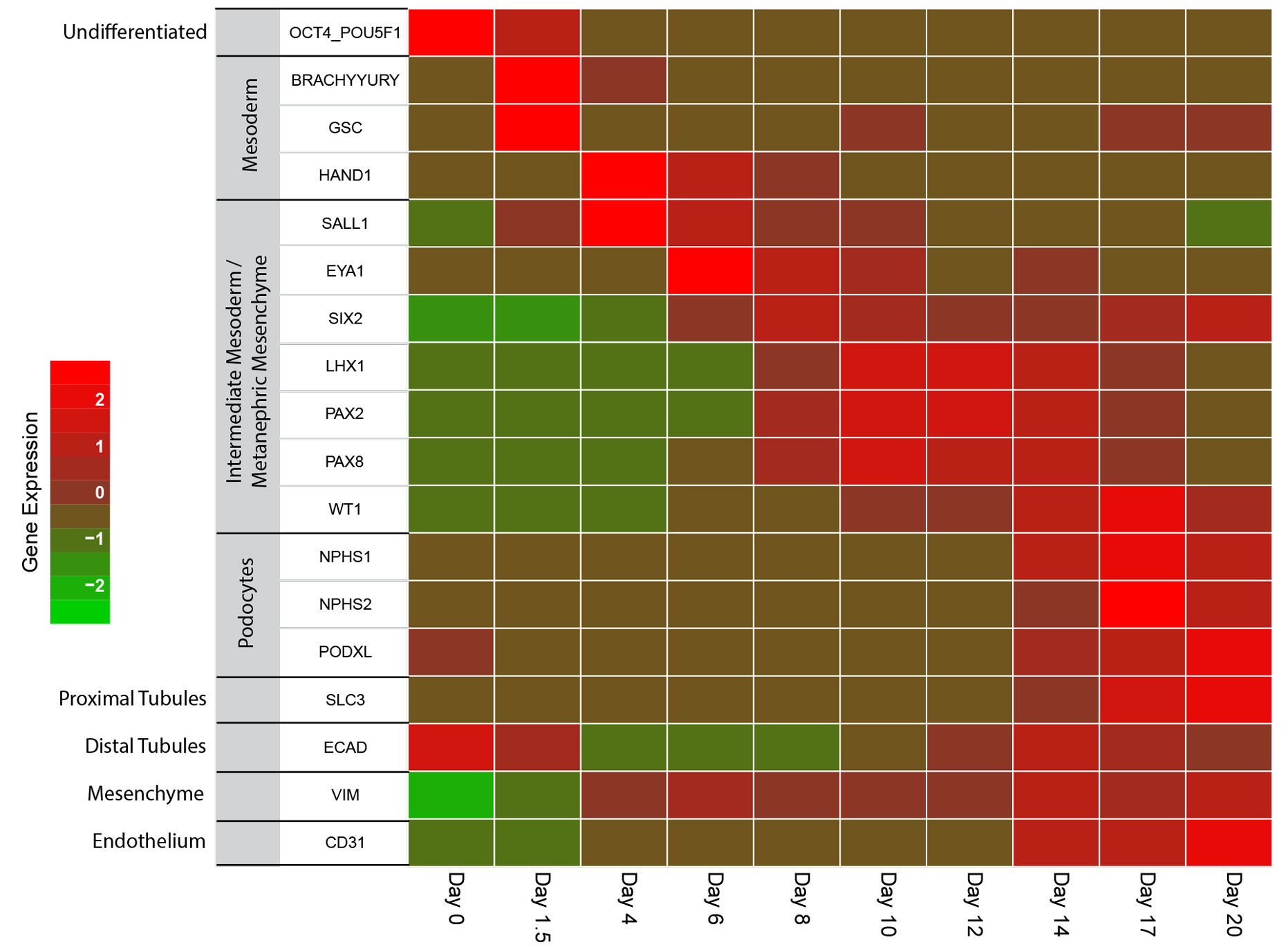
Figure 4. Kidney Organoids Grown Using the STEMdiff™ Kidney Organoid Kit Show the Expected Changes in Gene Expression During Differentiation
As organoids progress through the stages of differentiation to more mature renal cell types, gene expression shifts from markers of pluripotency (day 0) to the mesoderm (day 1.5 - 4) and to the intermediate mesoderm/metanephric mesenchyme by day 4 - 12. Markers of the podocytes, proximal and distal tubules, mesenchyme and endothelium are observable starting by day 14. Marker levels were assessed in four independent experiments by RT-qPCR and normalized to expression levels of undifferentiated cells.
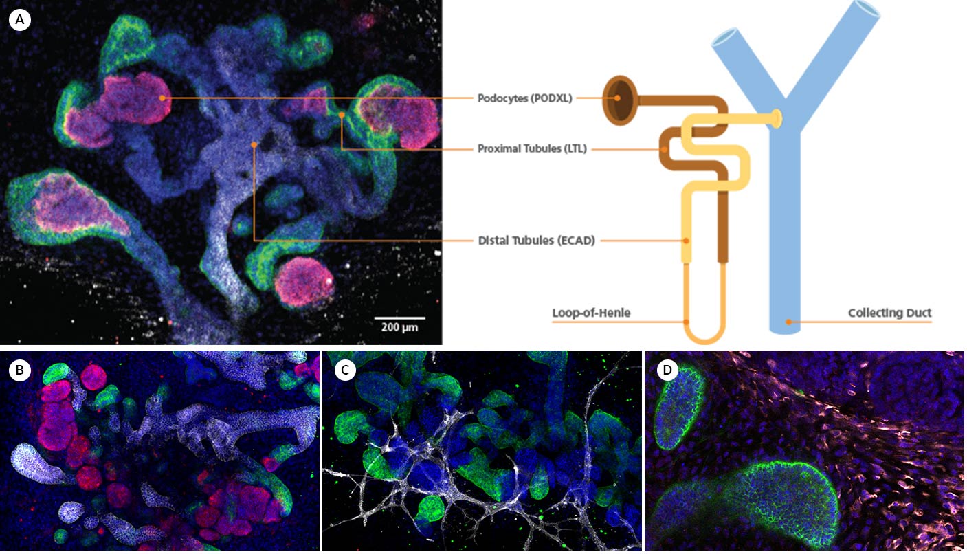
Figure 5. Kidney Organoids form Convoluted Tubular Structures with Typical Nephron-like Segmentation
(A) During differentiation, kidney organoids form convoluted tubular structures that resemble the structure and segmentation of the developing nephron. These organoids express markers of the (B) renal epithelium including podocalyxin (PODXL), lotus tetragonolobus lectin (LTL), and E-cadherin (ECAD), as well as markers of the (C) endothelium (platelet endothelial cell adhesion molecule, CD31), and (D) mesenchyme (vimentin, VIM; Meis homeobox family, MEIS1/2/3).

