STEMdiff™ Intestinal Organoid Kit
For extended culture and passaging, the kit components required for organoid maintenance can be purchased as STEMdiff™ Intestinal Organoid Growth Medium (Catalog #05145).
• Robust medium supports efficient differentiation of human ES and iPS cell lines to intestinal organoids
• Convenient model system that can be maintained long-term through passaging or cryopreserved for experimental flexibility
• Serum-free medium system optimized for low experimental variability
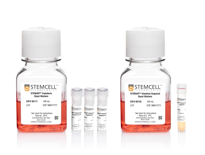
- STEMdiff™ Intestinal Organoid Kit (Catalog #05140)
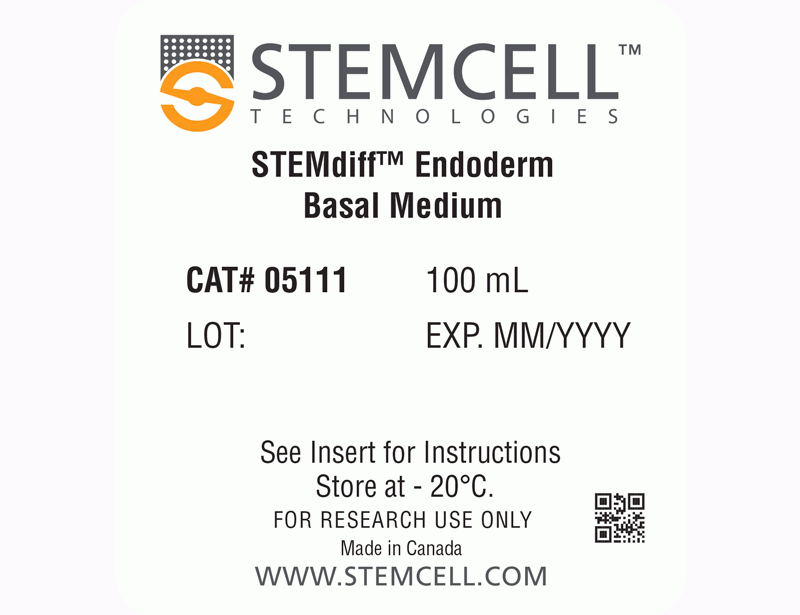
- STEMdiff™ Endoderm Basal Medium,100 mL
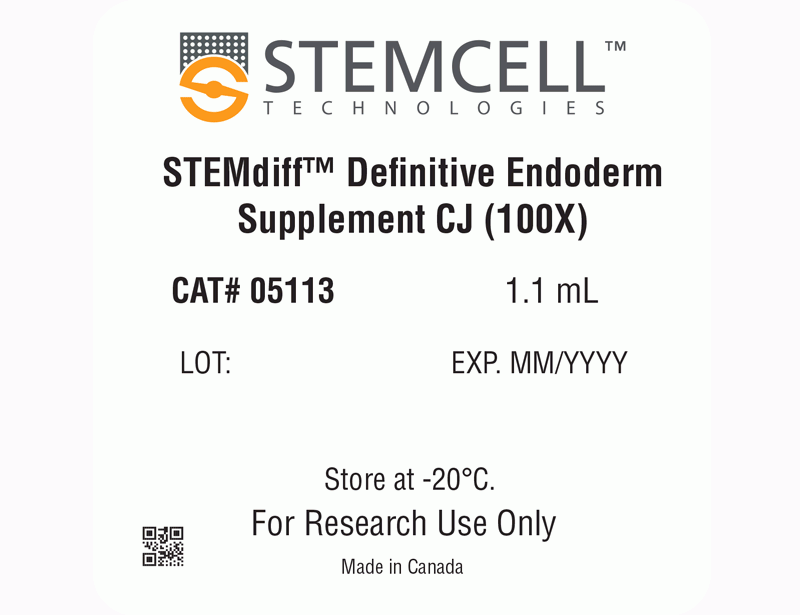
- STEMdiff™ Definitive Endoderm Supplement CJ, 1.1 mL
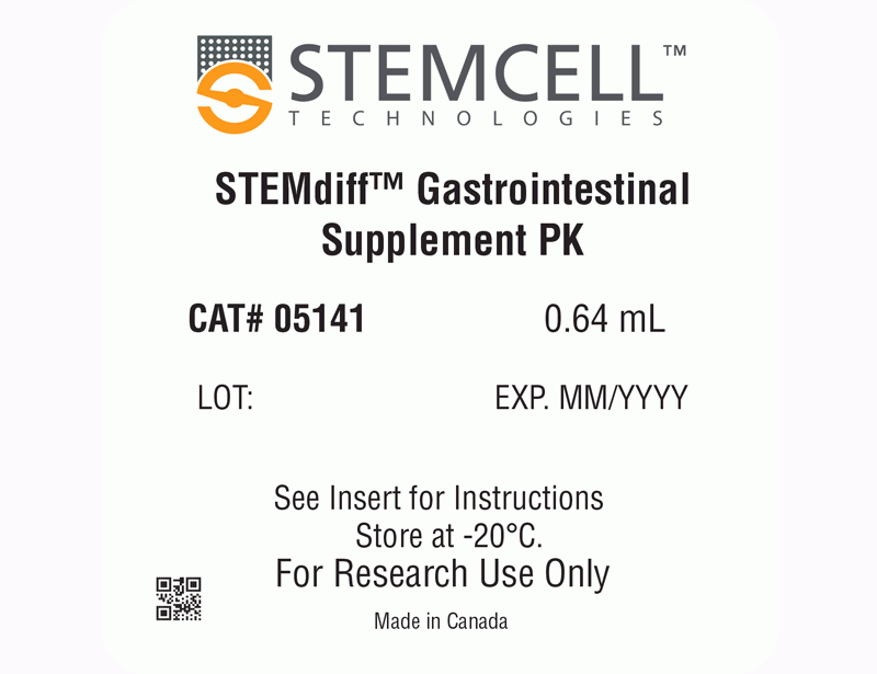
- STEMdiff™ Gastrointestinal Supplement PK, 0.64 mL
- STEMdiff™ Gastrointestinal Supplement UB, 0.64 mL
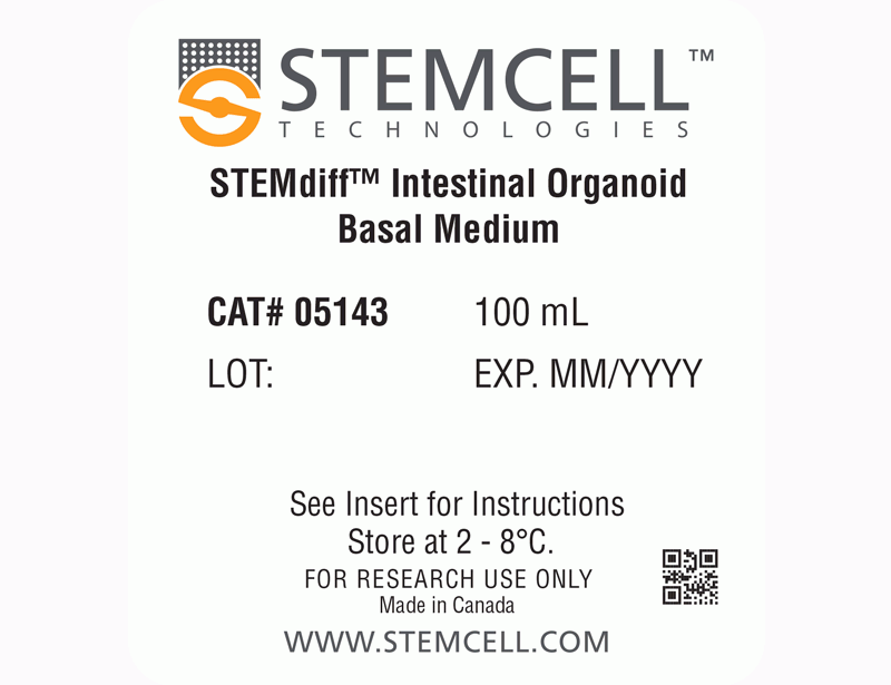
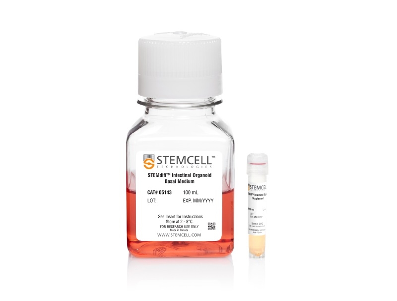
- STEMdiff™ Intestinal Organoid Basal Medium, 100 mL
- STEMdiff™ Intestinal Organoid Supplement, 2 mL
- STEMdiff™ Intestinal Organoid Growth Medium (Catalog #05145)
- STEMdiff™ Intestinal Organoid Basal Medium, 100 mL
- STEMdiff™ Intestinal Organoid Supplement, 2 mL
Data
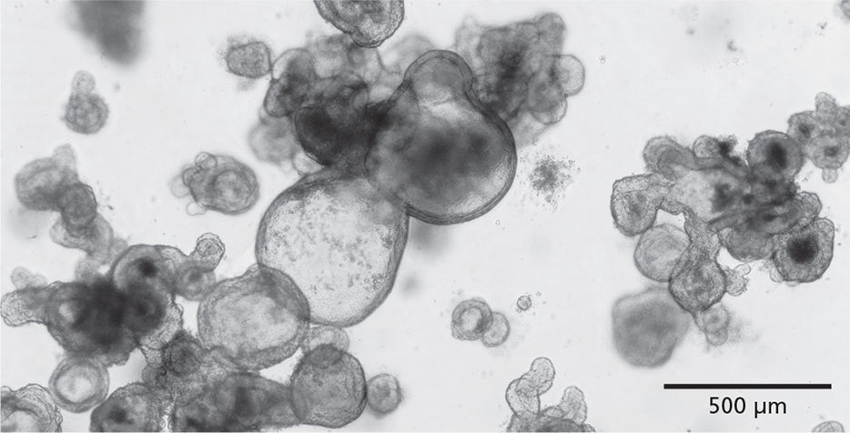
Figure 1. STEMdiff™ Intestinal Organoid Kit Enables the Growth of Intestinal Organoids from PSCs
STEMdiff™ Intestinal Organoid Kit facilitates directed differentiation of PSCs to form human small intestinal organoids. The organoids exhibit a polarized epithelial monolayer surrounding a hollow lumen and are associated with a mesenchymal cell population. Pictured are passage 3 human intestinal organoids generated using this kit.
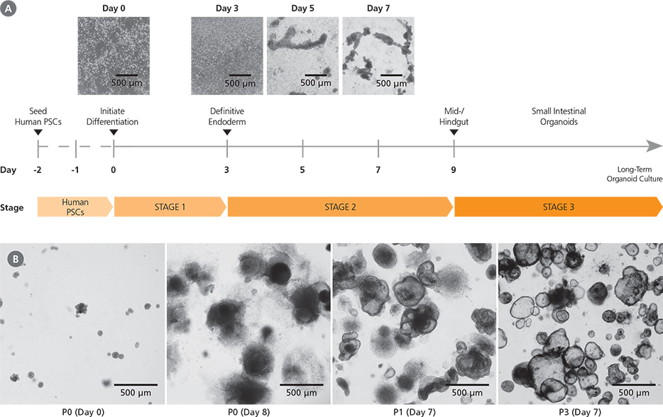
Figure 2. Generation of Human Intestinal Organoid Cultures Using the STEMdiff™ Intestinal Organoid Kit
(A) Human PSC cultures progress through a three-stage differentiation process to generate human intestinal organoids. By day 3 of the protocol, cultures exhibit characteristics typical of definitive endoderm and mid-/hindgut differentiation is initiated. During mid-/hindgut differentiation (days 5 - 9), cells form mid-/hindgut spheroids that are released from the cell monolayer into the culture medium. These spheroids are collected and embedded in extracellular matrix. (B) Embedded mid-/hindgut spheroids cultured in STEMdiff™ Intestinal Organoid Growth Medium mature into intestinal organoids (days in parentheses indicate days post-embedding in a given passage). Once established, intestinal organoids can be maintained and expanded in culture by passaging every 7 - 10 days. After multiple passages, the organoids generally exhibit less sinking within the matrix dome and a lower proportion of mesenchymal cells.

Figure 3. STEMdiff™ Intestinal Organoid Kit Supports Robust Differentiation and Expansion Across ESC and iPSC Lines
STEMdiff™ Intestinal Organoid Kit enables high-efficiency generation of intestinal organoids from both ESCs (H9, H7) and iPSCs (WLS-1C, STiPS-MOO1). (A) Organoids initiated from a variety of cell lines show efficient induction of definitive endoderm, measured by co-expression of FOXA2 and SOX17 on day 3 of differentiation. (B) Both ESCand iPSC-derived cultures demonstrate efficient spheroid formation upon mid-/hindgut induction. The total number of spheroids obtained per well in a given differentiation is shown. (C) Organoids cultured from either ESCs or iPSCs can be expanded and maintained over multiple passages. Shown is the total cell yield per passage. Organoids were passaged every 7 - 10 days with a split ratio between 1 in 2 and 1 in 4. Data points represent the mean of 3 biological replicates. Error bars throughout represent standard deviation of the mean.
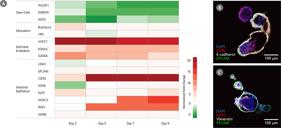
Figure 4. Characteristics of Mid-/Hindgut Spheroids Generated with STEMdiff™ Intestinal Organoid Kit
(A) Cultures differentiated using STEMdiff™ Intestinal Organoid Kit exhibit the expected markers during definitive endoderm and mid-/hindgut specification. During the protocol, gene expression patterns shift from pluripotency markers (day 0) to definitive endoderm markers by day 3 and those of the mid-/hindgut epithelium by day 9. Mid-/hindgut cultures (day 9) also express markers of the associated mesenchyme. Marker levels were assessed by RT-qPCR and normalized to expression levels for undifferentiatied H9 cells. (B) Mid-/hindgut spheroids (day 9) express markers of the intestinal epithelium (CDX2, E-cadherin, EPCAM). (C) Mid-/hindgut spheroids (day 9) also incorporate components of the associated mesenchyme (vimentin).
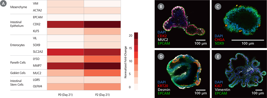
Figure 5. Intestinal Organoids Cultured In STEMdiff™ Intestinal Organoid Growth Medium Exhibit Features of the Intestinal Epithelium
(A) Differentiated PSC-derived intestinal organoids express markers of the intestinal epithelium and the associated mesenchyme. Marker levels were assessed by RT-qPCR and normalized to expression levels for undifferentiated H9 cells. (B,C) Intestinal organoids express markers of intestinal progenitor cells including CDX2 and the intestinal crypt marker SOX9. The organoids are composed of a polarized epithelium, visualized by the localization of EPCAM to the exterior (basolateral) surface of the organoids (B), and express markers typical of mature cell types including MUC2 (B: goblet cells) and CHGA (C: enteroendocrine cells). (D,E) Observation of desmin (D) and vimentin (E) in intestinal organoids demonstrates incorporation of mesenchymal cells in the organoid cultures, while KRT20 (D) and Ki67 (E) are markers of differentiated intestinal cells and putative intestinal stem cells, respectively. Images are digital cross-sections of whole-mount immunofluorescence-stained intestinal organoids at P28 (Day 7).
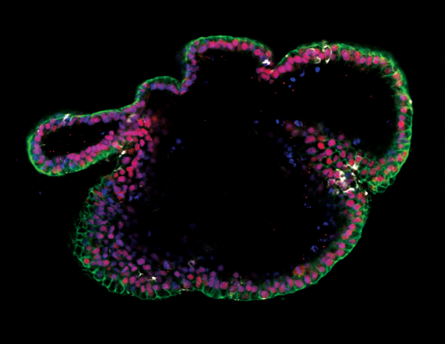
Figure 6. Generation of Intestinal Organoids from hPSCs Maintained in mTeSR™ Plus
Human ES (H9) cells were cultured with mTeSR™ Plus and directed to intestinal organoids using the STEMdiff™ Intestinal Organoid Kit. Image shows markers of the intestinal epithelium EpCAM (green) and CDX2 (red), and intestinal mesenchyme marker vimentin (white). Nuclei are counterstained with DAPI (blue).

