
STEMdiff™ Endothelial Differentiation Kit

⦁ No cell enrichment or sorting step required.
⦁ Superior hPSC-derived endothelial expansion compared to FBS-contain media.
- STEMdiff™ Endothelial Induction Medium Kit (Catalog #08005)
- STEMdiff™ Endothelial Induction Medium, 100 mL
- STEMdiff™ Endothelial Expansion Basal Medium, 120 mL
- STEMdiff™ Endothelial Expansion 5X Supplement, 30 mL
- Animal Component-Free Cell Attachment Substrate, 1 mL
| Document Type | 产品名称 | Catalog # | Lot # | 语言 |
|---|---|---|---|---|
| Product Information Sheet | STEMdiff™ Endothelial Differentiation Kit | 08005 | All | English |
| Safety Data Sheet 1 | STEMdiff™ Endothelial Differentiation Kit | 08005 | All | English |
| Safety Data Sheet 2 | STEMdiff™ Endothelial Differentiation Kit | 08005 | All | English |
| Safety Data Sheet 3 | STEMdiff™ Endothelial Differentiation Kit | 08005 | All | English |
| Safety Data Sheet 4 | STEMdiff™ Endothelial Differentiation Kit | 08005 | All | English |
Data
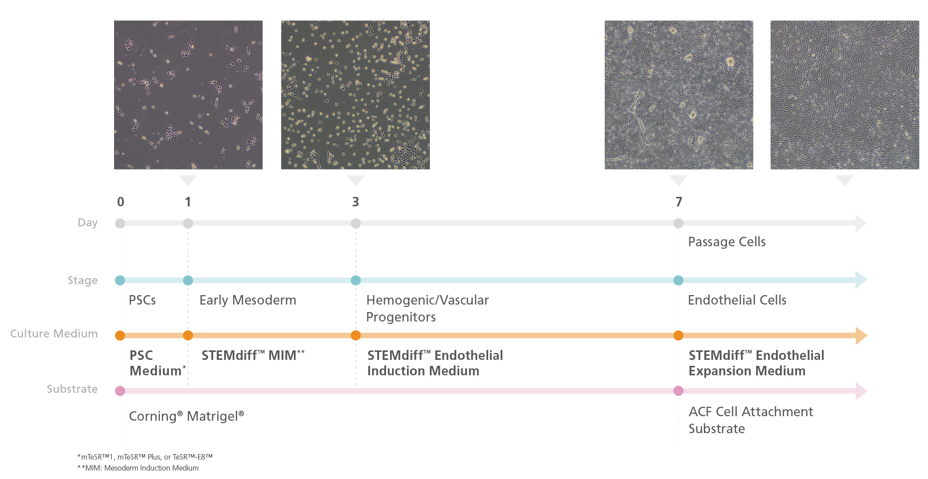
Figure 1. Schematic Workflow of Endothelial Induction Using the STEMdiff™ Endothelial Kit
In Phase 1, human embryonic stem (ES) or induced pluripotent stem (iPS) cells are cultured in a TeSR™ maintenance medium (mTeSR™ Plus, mTeSR™1, or TeSR™-E8™). On Day 1 (Phase 2) of the protocol, cells are ready for induction into early mesoderm progenitor cells by replacing TeSR™ medium with STEMdiff™ Mesodermal Induction Medium (MIM). By Day 3 (Phase 3), STEMdiff™ Mesoderm Induction Medium is replaced with STEMdiff™ Endothelial Induction Medium to derive endothelial cells. On Day 7, cells are passaged 5 - 6 times onto cultureware pre-coated with Animal Component-Free Cell Attachment Substrate in STEMdiff™ Endothelial Expansion Medium (Phase 4).
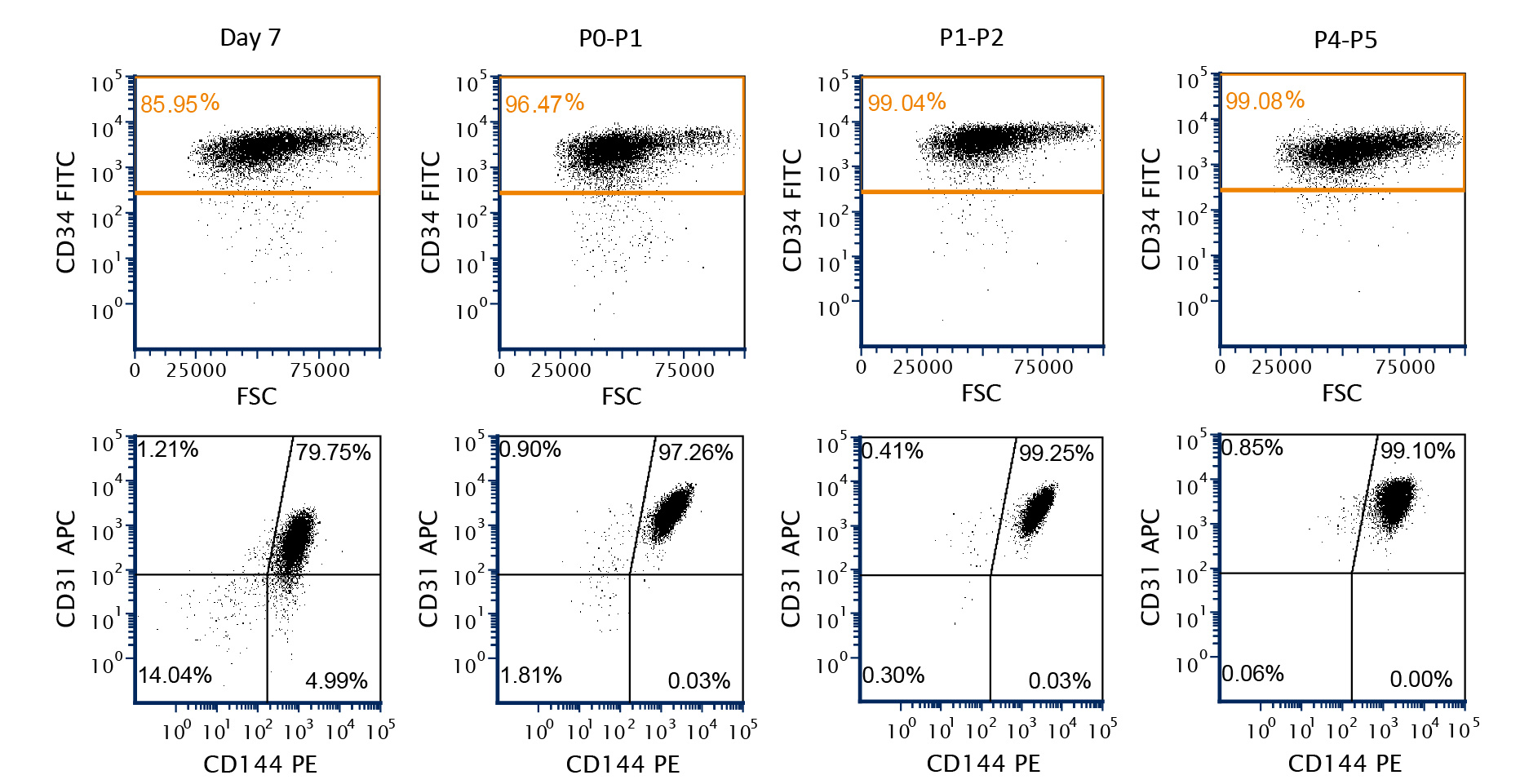
Figure 2. A Representative Flow Cytometric Analysis of Endothelial Marker Expression in hPSC-Derived Endothelial Cells
Human pluripotent stem cell (hPSC; H9 cell line)-derived endothelial cells were obtained at Day 7 using STEMdiff™ Endothelial Induction Medium. Greater than 85% of the cells were CD34+ and had high levels of CD31 and CD144 expression. With subsequent passages, the proportion of cells expressing endothelial markers (CD34+, CD31, and CD144) increased up to passage 5.
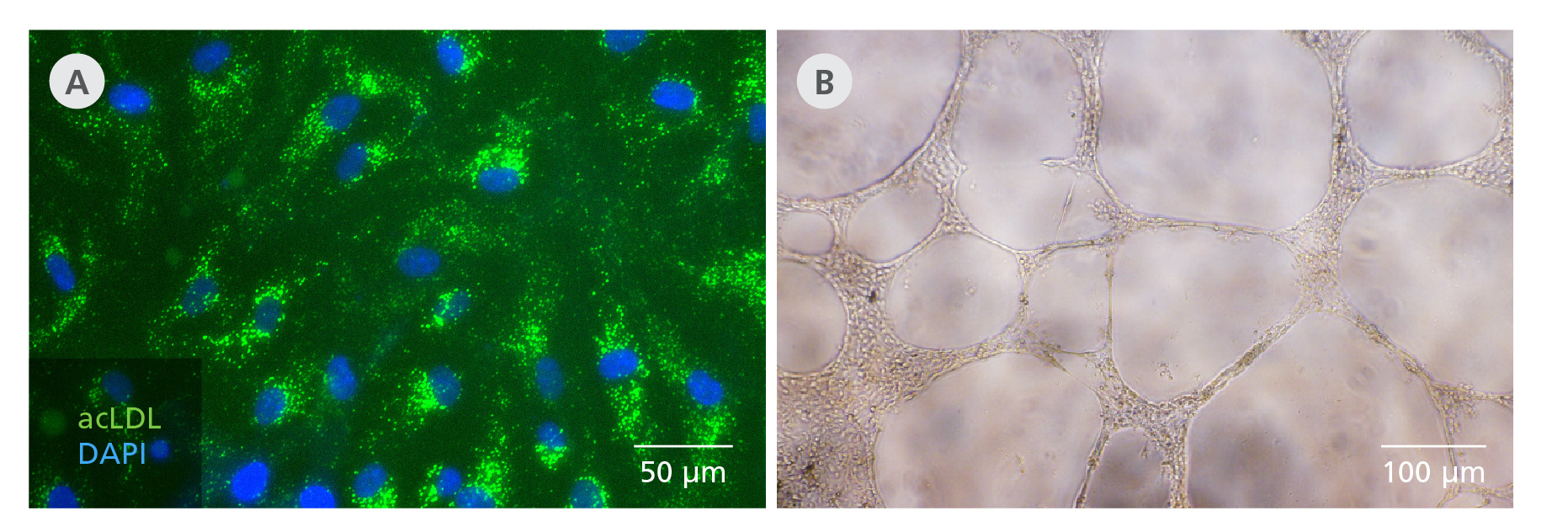
Figure 3. STEMdiff™ Endothelial Differentiation Kit Generates Functional hPSC-Derived Endothelial Cells
Endothelial cells generated from hPSCs (F016 cell line) using the STEMdiff™ Endothelial Differentiation Kit take up acetylated LDL when plated at 10,000 cells/cm2. Cells are able to form tubular networks in vitro in a tube formation assay when plated at 20,000 cells/well in a 96 well-plate for 24 hrs.
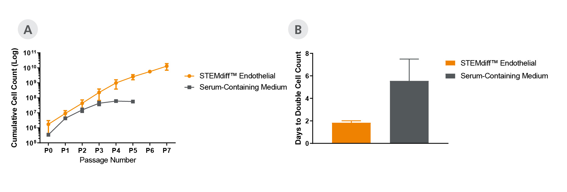
Figure 4. Endothelial Cells Expand Faster in STEMdiff™ Endothelial Expansion Medium Compared to Serum-Containing Medium
STEMdiff™ Endothelial Expansion Medium (A) sustains expansion rate in later passages and leads to (B) superior expansion of hPSC (C1 cell line)-derived endothelial cells when compared to serum-containing medium.
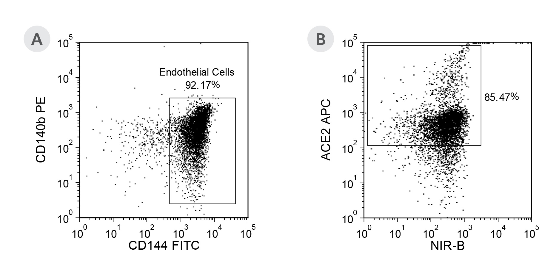
Figure 5. hPSC-Derived Endothelial Cells Generated Using the STEMdiff™ Endothelial Differentiation Kit Express High Levels of ACE2
(A) hPSC (C1 cell line)-derived endothelial cells were generated using the STEMdiff™ Endothelial Differentiation Kit and expanded in STEMdiff™ Endothelial Expansion Medium for 6 passages at 10,000 cells/cm2. (B) The cells were then analyzed for expression of angiotensin-converting enzyme 2 (ACE2). 85% of cells expressed high levels of ACE2.