
PneumaCult™-ALI-S Medium
Together, PneumaCult™-ALI-S Medium and PneumaCult™-Ex Plus Medium (#05040) constitute a fully integrated BPE-free culture system for in vitro human small airway modeling. This robust and defined system is a valuable tool for basic respiratory research, toxicity studies, and drug development.
• PneumaCult™-ALI-S is serum-free and BPE-free to minimize variability
• PneumaCult™-ALI-S and PneumaCult™-Ex Plus constitute a complete, optimized system for expansion, maintenance, and differentiation of HSAEC
- PneumaCult™-ALI-S Basal Medium, 450 mL
- PneumaCult™-ALI-S Supplement (10X), 50 mL
- PneumaCult™-ALI-S Maintenance Supplement (100X), 5 x 1 mL
| Document Type | 产品名称 | Catalog # | Lot # | 语言 |
|---|---|---|---|---|
| Product Information Sheet | PneumaCult™-ALI-S Medium | 05050 | All | English |
| Special Protocol | PneumaCult™-ALI-S Medium | 05050 | All | English |
| Safety Data Sheet 1 | PneumaCult™-ALI-S Medium | 05050 | All | English |
| Safety Data Sheet 2 | PneumaCult™-ALI-S Medium | 05050 | All | English |
| Safety Data Sheet 3 | PneumaCult™-ALI-S Medium | 05050 | All | English |
Data
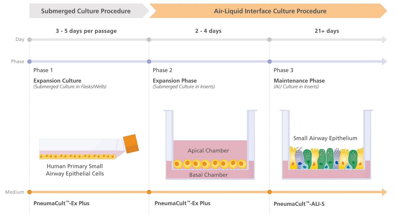
Figure 1. Overview of the PneumaCult™ Culture System for Small Airway Research
Expansion of human small airway epithelial cells (HSAEC) in submerged culture is performed with PneumaCult™-Ex Plus Medium. During the early Expansion Phase of the air-liquid interface (ALI) culture procedure, PneumaCult™-Ex Plus is applied to the apical and basal chambers. Upon reaching confluence, the culture is air-lifted by removing the culture medium from both chambers, and PneumaCult™-ALI-S is added to the basal chamber only. Differentiation into a mucociliary epithelium is obtained following 21+ days of incubation and can be maintained for more than one year.
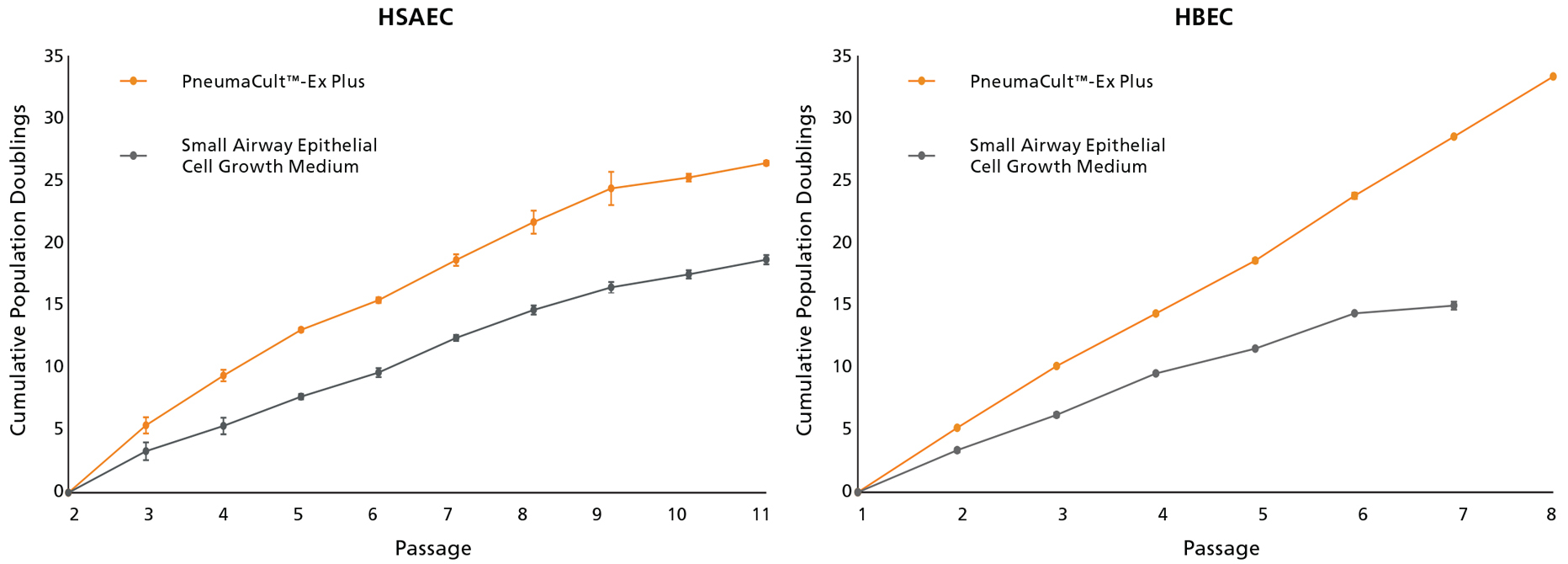
Figure 2. HSAEC and HBEC Grow at a Higher Rate During Expansion When Cultured in PneumaCult™-Ex Plus Medium
Human small airway epithelial cells (HSAEC) and human bronchial epithelial cells (HBEC) cultured in PneumaCult™-Ex Plus Medium exhibited higher proliferation rate at every passage compared with cells cultured in Small Airway Epithelial Cell Growth Medium. Cryopreserved HSAEC were obtained commercially at passage 2 (P2) while HBEC were obtained at P1.
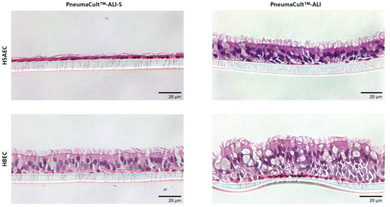
Figure 3. HSAEC Cultured at the ALI Using PneumaCult™-ALI-S Medium Differentiate to Form a Morphology Representative of the Small Airway Epithelium
Hematoxylin and eosin (H&E) staining of HSAEC and HBEC cultured in PneumaCult™-ALI-S or PneumaCult™-ALI Medium at P3, after 28 days. HSAEC differentiated at the ALI in PneumaCult™-ALI-S formed a thin, cuboidal epithelial layer representative of the in vivo small airway epithelium while HBEC differentiated in PneumaCult™-ALI formed a pseudostratified epithelium resembling the in vivo bronchial epithelium. The ALI cultures were fixed, paraffin-embedded, sectioned, and stained with H&E. All images were taken using a 40X objective. Insert membrane was 10 μm in thickness. Scale bar = 20 μm.
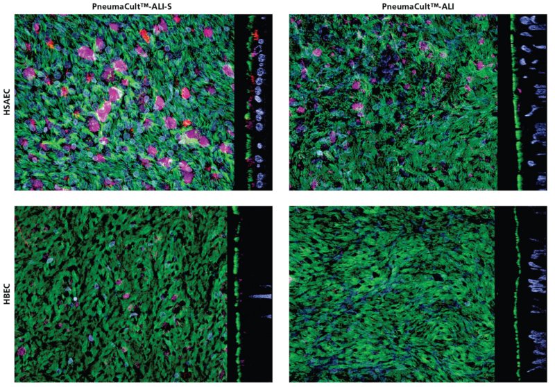
Figure 4. Small Airway Epithelium Markers Were Detected in HSAEC Cultured in PneumaCult™-ALI-S Medium
Confocal images of whole mount immunostained ALI cultures showing HSAEC and HBEC cultured in PneumaCult™-ALI-S or PneumaCult™-ALI Medium at P3, after 28 days. The ALI cultures were fixed and stained with antibodies for ciliated cells (AC-tubulin; green), club cells (SCGB1A1; magenta), and secretory protein (SCGB3A2; red). The nuclei were counterstained with DAPI (blue). Small airway epithelium markers, SCGB1A1 and SCGB3A2, were detected at higher levels in HSAEC cultured in PneumaCult™-ALI-S compared with HSAEC cultured in PneumaCult™-ALI and HBEC cultured in either PneumaCult™-ALI-S or PneumaCult™-ALI. All images were taken using a 63X objective.
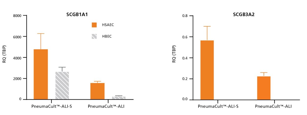
Figure 5. Relative Expression of Small Airway Epithelium Markers by qPCR Were Detected at Higher Levels in HSAEC Cultured in PneumaCult™-ALI-S Medium Compared with HSAEC Cultured in PneumaCult™-ALI
HSAEC and HBEC cultured in PneumaCult™-ALI-S or PneumaCult™-ALI Medium at P3. After 28-days of differentiation, the ALI cultures were analysed for small airway epithelium markers, SCGB1A1 and SCGB3A2. Gene of Interest expression was normalized to housekeeping gene, TBP, and expressed as relative quantity (RQ). Relative expression of SCGB1A1 and SCGB3A2 was higher in HSAEC cultured in PneumaCult™-ALI-S Medium compared with HSAEC cultured in PneumaCult™-ALI and HBEC cultured in either PneumaCult™-ALI-S or PneumaCult™-ALI. Relative expression of SCGB3A2 was not detectable in HBEC cultured in either PneumaCult™-ALI or PneumaCult™-ALI-S.