NeuroCult™ Proliferation Supplement (Mouse & Rat)
Supplement for expansion of mouse and rat neural stem and progenitor cells
概要
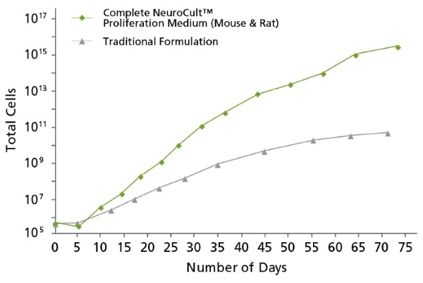
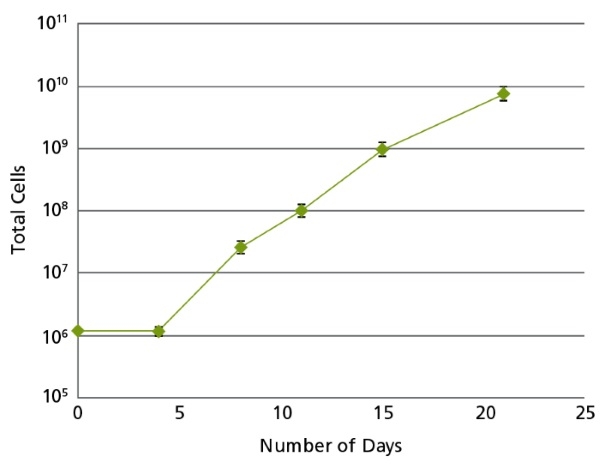
NeuroCult™ Proliferation Supplement (Mouse & Rat) is a standardized, serum-free supplement for the culture of mouse and rat neural stem and progenitor cells. NeuroCult™ Proliferation Supplement (Mouse & Rat) is designed to be combined with NeuroCult™ Basal Medium (Mouse & Rat; Catalog #05700) and appropriate cytokines (not included). NeuroCult™ Proliferation Supplement (Mouse & Rat) is optimized to maintain mouse and rat neural stem cells in culture for extended periods of time without the loss of their self-renewal, proliferation, or differentiation potential. This supplement is also a component of NeuroCult™ Proliferation Kit (Mouse & Rat; Catalog #05702).
NOTE: When preparing Complete NeuroCult™ Proliferation Medium, addition of Human Recombinant EGF (Catalog #78006.1) is required. When culturing cells obtained from adult mouse or rat, Human Recombinant bFGF (Catalog #78003.1) and Heparin Solution (Catalog #07980) are also required.
Brain Tumor Stem Cells, Neural Stem and Progenitor Cells
Cell Culture, Colony Assay, Expansion, Functional Assay, Spheroid Culture
Cancer Research, Disease Modeling, Neuroscience, Stem Cell Biology
数据及文献
Publications (117)
Neuroscience Letters 2017 MAR
Recombinant insulin-like growth factor binding protein-4 inhibits proliferation and promotes differentiation of neural progenitor cells
Niu H et al.
Abstract
Insulin-like growth factor (IGF) is involved in regulating many processes during neural development, and IGF binding protein-4 (IGFBP4) functions as a modulator of IGF actions or in an IGF-independent manner (e.g., via inhibiting Wnt/β-catenin signaling). In the present study, neural progenitor cells (NPCs) were isolated from the forebrain of newborn mice to investigate effects of IGFBP4 on the proliferation and differentiation of NPCs. The proliferation of NPCs was evaluated using Cell Counting Kit-8 (CCK-8) after treatment with or without IGFBP4 as well as blockers of IGF-IR and β-catenin. Phosphorylation levels of Akt, Erk1, 2 and p38 were analyzed by Western blotting. The differentiation of NPCs was evaluated using immunofluorescence and Western blotting. It was shown that exogenous IGFBP4 significantly inhibited the proliferation of NPCs and it did not induce a more pronounced inhibition of cell proliferation after blockade of IGF-IR but it did after antagonism of β-catenin. Akt phosphorylation was significantly decreased and phosphorylation levels of Erk1, 2 and p38 were not significantly changed in IGFBP4-treated NPCs. Excessive IGFBP4 significantly promoted NPCs to differentiate into astrocytes and neurons. These data suggested that exogenous IGFBP4 inhibits proliferation and promotes differentiation of neural progenitor cells mainly through IGF-IR signaling pathway.
Journal of neuroimmune pharmacology : the official journal of the Society on NeuroImmune Pharmacology 2017 JUN
Cathepsin B Improves ß-Amyloidosis and Learning and Memory in Models of Alzheimer's Disease.
Embury CM et al.
Abstract
Amyloid-ß (Aß) precursor protein (APP) metabolism engages neuronal endolysosomal pathways for Aß processing and secretion. In Alzheimer's disease (AD), dysregulation of APP leads to excess Aß and neuronal dysfunction; suggesting that neuronal APP/Aß trafficking can be targeted for therapeutic gain. Cathepsin B (CatB) is a lysosomal cysteine protease that can lower Aß levels. However, whether CatB-modulation of Aß improves learning and memory function deficits in AD is not known. To this end, progenitor neurons were infected with recombinant adenovirus expressing CatB and recovered cell lysates subjected to proteomic analyses. The results demonstrated Lamp1 deregulation and linkages between CatB and the neuronal phagosome network. Hippocampal injections of adeno-associated virus expressing CatB reduced Aß levels, increased Lamp1 and improved learning and memory. The findings were associated with the emergence of c-fos + cells. The results support the idea that CatB can speed Aß metabolism through lysosomal pathways and as such reduce AD-associated memory deficits.
Nature genetics 2017 JAN
Qki deficiency maintains stemness of glioma stem cells in suboptimal environment by downregulating endolysosomal degradation.
Shingu T et al.
Abstract
Stem cells, including cancer stem cells (CSCs), require niches to maintain stemness, yet it is unclear how CSCs maintain stemness in the suboptimal environment outside their niches during invasion. Postnatal co-deletion of Pten and Trp53 in mouse neural stem cells (NSCs) leads to the expansion of these cells in their subventricular zone (SVZ) niches but fails to maintain stemness outside the SVZ. We discovered that Qki is a major regulator of NSC stemness. Qk deletion on a Pten-/-; Trp53-/- background helps NSCs maintain their stemness outside the SVZ in Nes-CreERT2; QkL/L; PtenL/L; Trp53L/L mice, which develop glioblastoma with a penetrance of 92% and a median survival time of 105 d. Mechanistically, Qk deletion decreases endolysosome-mediated degradation and enriches receptors essential for maintaining self-renewal on the cytoplasmic membrane to cope with low ligand levels outside niches. Thus, downregulation of endolysosome levels by Qki loss helps glioma stem cells (GSCs) maintain their stemness in suboptimal environments outside their niches.
Stem Cell Research & Therapy 2017 DEC
CCL11 promotes migration and proliferation of mouse neural progenitor cells
Wang F et al.
Abstract
BACKGROUND Neonatal hypoxia-ischemia induces massive brain damage during the perinatal period, resulting in long-term consequences to central nervous system structural and functional maturation. Although neural progenitor cells (NPCs) migrate through the parenchyma and home in to injury sites in the rodent brain, the molecular mechanisms are unknown. We examined the role of chemokines in mediating NPC migration after neonatal hypoxic-ischemic brain injury. METHODS Nine-day-old mice were exposed to a 120-minute hypoxia following unilateral carotid occlusion. Chemokine levels were quantified in mouse brain extract. Migration and proliferation assays were performed using embryonic and infant mouse NPCs. RESULTS The neonatal hypoxic-ischemic brain injury resulted in an ipsilateral lesion, which was extended to the cortical and striatal areas. NPCs migrated toward an injured area, where a marked increase of CC chemokines was detected. In vitro studies showed that incubation of NPCs with recombinant mouse CCL11 promoted migration and proliferation. These effects were partly inhibited by a CCR3 antagonist, SB297006. CONCLUSIONS Our data implicate an important effect of CCL11 for mouse NPCs. The effective activation of NPCs may offer a promising strategy for neuroregeneration in neonatal hypoxic-ischemic brain injury.
Journal of neuroinflammation 2016 SEP
Metabolic determinants of the immune modulatory function of neural stem cells.
Drago D et al.
Abstract
BACKGROUND Neural stem cells (NSCs) display tissue trophic and immune modulatory therapeutic activities after transplantation in central nervous system disorders. The intercellular interplay between stem cells and target immune cells is increased in NSCs exposed to inflammatory cues. Here, we hypothesize that inflammatory cytokine signalling leads to metabolic reprogramming of NSCs regulating some of their immune modulatory effects. METHODS NSC lines were prepared from the subventricular zone (SVZ) of 7-12-week-old mice. Whole secretome-based screening and analysis of intracellular small metabolites was performed in NSCs exposed to cocktails of either Th1-like (IFN-γ, 500 U/ml; TNF-α, 200 U/ml; IL-1β, 100 U/ml) or Th2-like (IL-4, IL-5 and IL-13; 10 ng/ml) inflammatory cytokines for 16 h in vitro. Isotopologues distribution of arginine and downstream metabolites was assessed by liquid chromatography/mass spectrometry in NSCs incubated with U-(13)C6 L-arginine in the presence or absence of Th1 or Th2 cocktails (Th1 NSCs or Th2 NSCs). The expression of arginase I and II was investigated in vitro in Th1 NSCs and Th2 NSCs and in vivo in the SVZ of mice with experimental autoimmune encephalomyelitis, as prototypical model of Th1 cell-driven brain inflammatory disease. The effects of the inflammatory cytokine signalling were studied in NSC-lymph node cells (LNC) co-cultures by flow cytometry-based analysis of cell proliferation following pan-arginase inhibition with N(ω)-hydroxy-nor-arginine (nor-NOHA). RESULTS Cytokine-primed NSCs showed significantly higher anti-proliferative effect in co-cultures vs. control NSCs. Metabolomic analysis of intracellular metabolites revealed alteration of arginine metabolism and increased extracellular arginase I activity in cytokine-primed NSCs. Arginase inhibition by nor-NOHA partly rescued the anti-proliferative effects of cytokine-primed NSCs. CONCLUSIONS Our work underlines the use of metabolic profiling as hypothesis-generating tools that helps unravelling how stem cell-mediated mechanisms of tissue restoration become affected by local inflammatory responses. Among different therapeutic candidates, we identify arginase signalling as novel metabolic determinant of the NSC-to-immune system communication.
Scientific reports 2016 NOV
Sex-Specific Effects of Testosterone on the Sexually Dimorphic Transcriptome and Epigenome of Embryonic Neural Stem/Progenitor Cells.
Bramble MS et al.
Abstract
The mechanisms by which sex differences in the mammalian brain arise are poorly understood, but are influenced by a combination of underlying genetic differences and gonadal hormone exposure. Using a mouse embryonic neural stem cell (eNSC) model to understand early events contributing to sexually dimorphic brain development, we identified novel interactions between chromosomal sex and hormonal exposure that are instrumental to early brain sex differences. RNA-sequencing identified 103 transcripts that were differentially expressed between XX and XY eNSCs at baseline (FDR%=%0.10). Treatment with testosterone-propionate (TP) reveals sex-specific gene expression changes, causing 2854 and 792 transcripts to become differentially expressed on XX and XY genetic backgrounds respectively. Within the TP responsive transcripts, there was enrichment for genes which function as epigenetic regulators that affect both histone modifications and DNA methylation patterning. We observed that TP caused a global decrease in 5-methylcytosine abundance in both sexes, a transmissible effect that was maintained in cellular progeny. Additionally, we determined that TP was associated with residue-specific alterations in acetylation of histone tails. These findings highlight an unknown component of androgen action on cells within the developmental CNS, and contribute to a novel mechanism of action by which early hormonal organization is initiated and maintained.
View All Publications