
NeuroCult™ Neuronal Plating Medium
• Tested for compatibility with BrainPhys™ Neuronal Medium
| Document Type | 产品名称 | Catalog # | Lot # | 语言 |
|---|---|---|---|---|
| Product Information Sheet | NeuroCult™ Neuronal Plating Medium | 05713 | All | English |
| Safety Data Sheet | NeuroCult™ Neuronal Plating Medium | 05713 | All | English |
Data
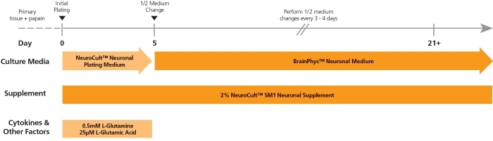
Figure 1. Protocol for Plating and Culturing Primary Neurons with the SM1 Culture System
Primary rodent tissue dissociated in papain was plated in NeuroCult™ Neuronal Plating Medium, supplemented with NeuroCult™ SM1 Neuronal Supplement, L-Glutamine, and L-Glutamic Acid. On day 5, primary neurons were transitioned to BrainPhys™ Neuronal Medium, supplemented with NeuroCult™ SM1 Neuronal Supplement, by performing half-medium changes every 3 - 4 days.
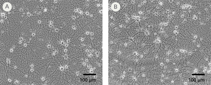
Figure 2. The SM1 Culture System Supports Long-Term Culture of Rodent Neurons
Primary E18 rat cortical neurons were cultured in the SM1 Culture System. A large number of viable neurons are visible after (A) 21 and (B) 35 days, as demonstrated by their bright neuronal cell bodies, and extensive neurite outgrowth and branching. Neurons are evenly distributed over the culture surface with minimal cell clumping.
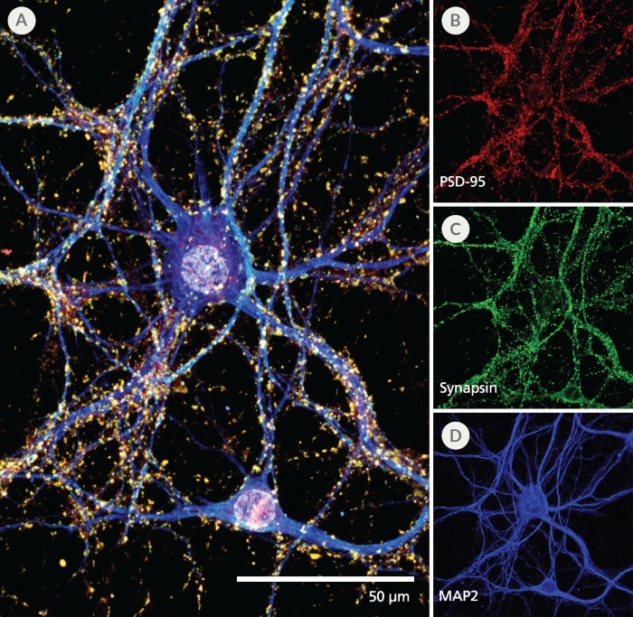
Figure 3. Pre- and Post-Synaptic Markers are Expressed in Rodent Neurons Cultured in the SM1 Culture System
Primary E18 rat cortical neurons were cultured in the SM1 Culture System. At 21 DIV, neurons are phenotypically mature, as indicated by the presence of an extensive dendritic arbor, and appropriate expression and localization of pre-synaptic synapsin (A,C; green) and post-synaptic PSD-95 (A,B; red) markers. Synapsin is concentrated in discrete puncta distributed along the somata and dendritic processes, as defined by the dendritic marker MAP2 (A,D; blue).
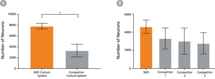
Figure 4. The SM1 Culture System Supports Increased Cell Survival
(A) Primary E18 rat cortical neurons were cultured in the SM1 Culture System or a Competitor Culture System for 21 days. Neurons cultured in the SM1 Culture System have a significantly higher number of viable cells compared to the competitor culture system (n = 4; mean ± 95% CI; *p < 0.05). (B) Primary E18 rat cortical neurons were cultured in Neurobasal® supplemented with NeuroCult™ SM1 Neuronal Supplement (SM1) or competitor B27-like supplements (Competitor 1,2,3) for 21 days. Cultures supplemented with NeuroCult™ SM1 Neuronal Supplement have an equal number of neurons compared to competitor-supplemented cultures. Bars represent standard error of mean.