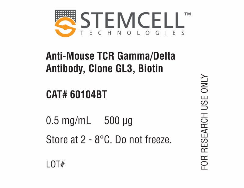
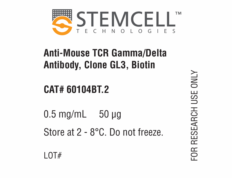
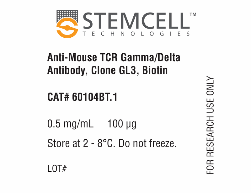
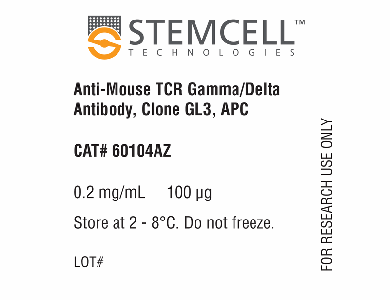
Anti-Mouse TCR Gamma/Delta Antibody, Clone GL3

Hamster (Armenian) monoclonal IgG2 antibody against mouse T Cell receptor gamma/delta
概要
The GL3 antibody reacts with the δ chain of the murine T cell receptor γ/δ (TCRγ/δ or TCR gamma/delta), a subtype of the TCR involved in the recognition of both peptide and lipid antigens. TCRγ/δ comprises a heterodimer (~ 80 kDa in humans) of disulfide-linked γ and δ subunits that associates with CD3 on the cell surface. It is a member of the immunoglobulin superfamily. TCRγ/δ is expressed on a subpopulation of T cells in the circulation but may be found on up to 50% of the T cells in epithelial cell-rich tissues. These cells have roles in oral and tumor-associated tolerance as well as autoimmune disease and have been described as a link between the adaptive and innate immune responses. Once activated, they secrete effector cytokines in a subtype- and context-specific manner. Most γ/δ T cells are CD4-/CD8-, though some express CD8. The GL3 antibody recognizes an epitope in the constant region of the δ chain and can reportedly activate TCRγ/δ+ cells.
Subtype
Primary Antibodies
Target Antigen
T Cell Receptor Gamma/Delta
Alternative Names
Gamma/Delta TCR , gdTCR, TCRgd, TCR γ/δ, T cell receptor delta chain, T cell receptor gamma chain, T cell receptor γ/δ, T3D, T3G
Reactive Species
Mouse
Conjugation
Alexa Fluor 488, APC, Biotin, FITC, PE, Unconjugated
Host Species
Hamster
Cell Type
T Cells, T Cells, Other Subsets
Species
Mouse
Application
Cell Isolation, Depletion, Flow Cytometry, Functional Assay, Immunofluorescence, Immunohistochemistry, Immunoprecipitation, Neutralization and Blocking
Area of Interest
Immunology
Clone
GL3
Gene ID
110066/110067
Isotype
IgG2, kappa
数据及文献

Data

Data for Unconjugated

Figure 1. Data for Unconjugated

(A) Flow cytometry analysis of C57BL/6 mouse lymph node cells labeled with Anti-Mouse TCR Gamma/Delta Antibody, Clone GL3, followed by an anti-hamster (Armenian) IgG antibody, FITC and Anti-Mouse CD3e Antibody, Clone 145-2C11, APC (Catalog #60015AZ).
Data for Alexa Fluor® 488-Conjugated

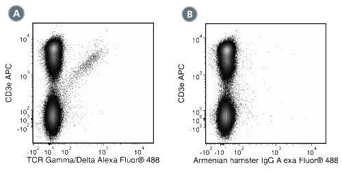
Figure 2. Data for Alexa Fluor® 488-Conjugated

(A) Flow cytometry analysis of C57BL/6 mouse lymph node cells labeled with Anti-Mouse TCR Gamma/Delta Antibody, Clone GL3, Alexa Fluor® 488 and Anti-Mouse CD3e Antibody, Clone 145-2C11, APC (Catalog #60015AZ).
Data for APC-Conjugated

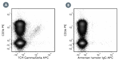
Figure 3. Data for APC-Conjugated

(A) Flow cytometry analysis of C57BL/6 mouse lymph node cells labeled with Anti-Mouse TCR Gamma/Delta Antibody, Clone GL3, APC and Anti-Mouse CD3e Antibody, Clone 145-2C11, PE (Catalog #60015PE).
Data for Biotin-Conjugated

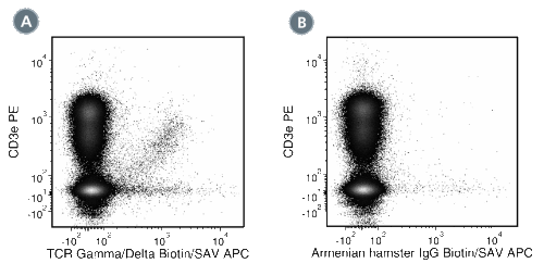
Figure 4. Data for Biotin-Conjugated

(A) Flow cytometry analysis of C57BL/6 mouse lymph node cells labeled with Anti-Mouse TCR Gamma/Delta Antibody, Clone GL3, Biotin followed by streptavidin (SAV) APC and Anti-Mouse CD3e Antibody, Clone 145-2C11, PE (Catalog #60015PE).
Data for FITC-Conjugated

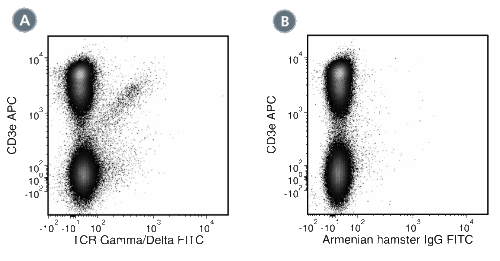
Figure 5. Data for FITC-Conjugated

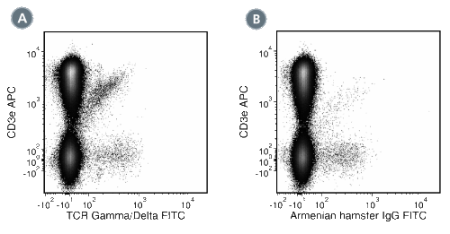
(A) Flow cytometry analysis of C57BL/6 mouse lymph node cells labeled with Anti-Mouse TCR Gamma/Delta Antibody, Clone GL3, FITC and Anti-Mouse CD3e Antibody, Clone 145-2C11, APC (Catalog #60015AZ).
Data for PE-Conjugated

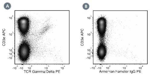
Figure 6. Data for PE-Conjugated

(A) Flow cytometry analysis of C57BL/6 mouse lymph node cells labeled with Anti-Mouse TCR Gamma/Delta Antibody, Clone GL3, PE and Anti-Mouse CD3e Antibody, Clone 145-2C11, APC (Catalog #60015AZ).

Top