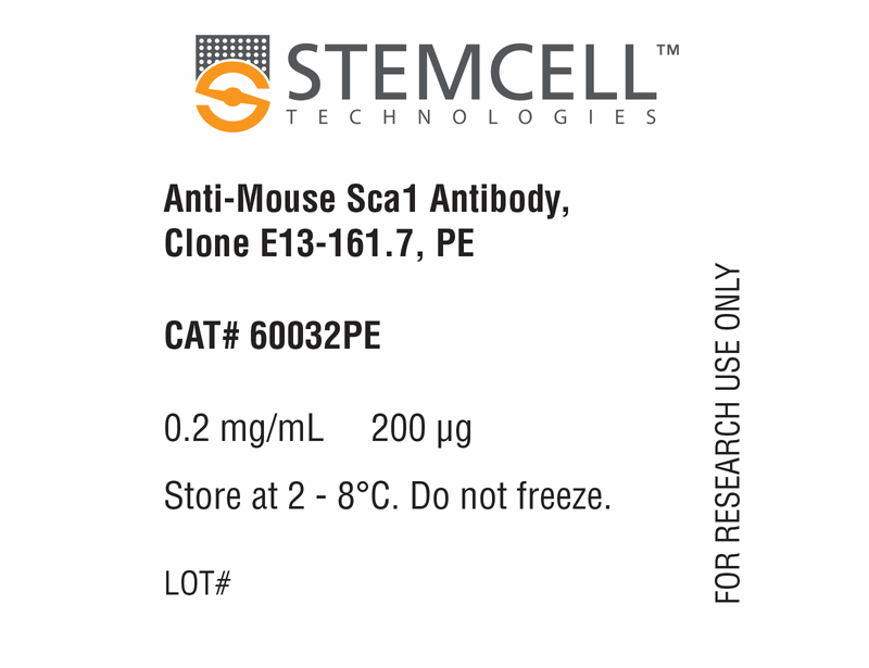
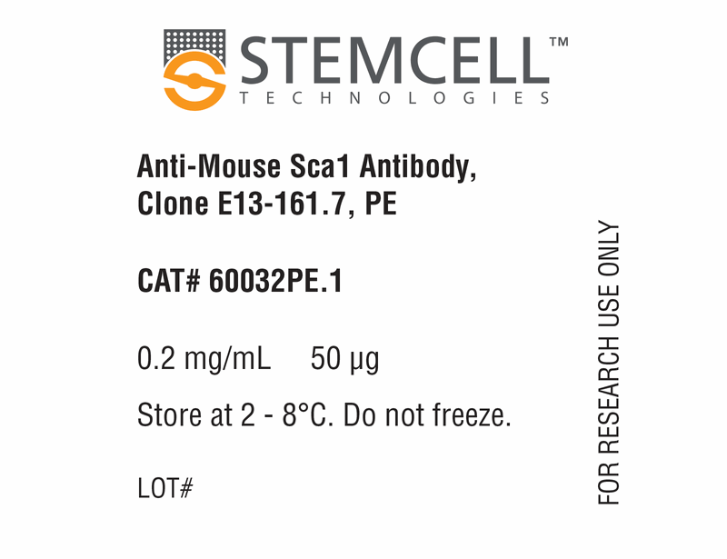
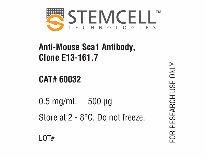
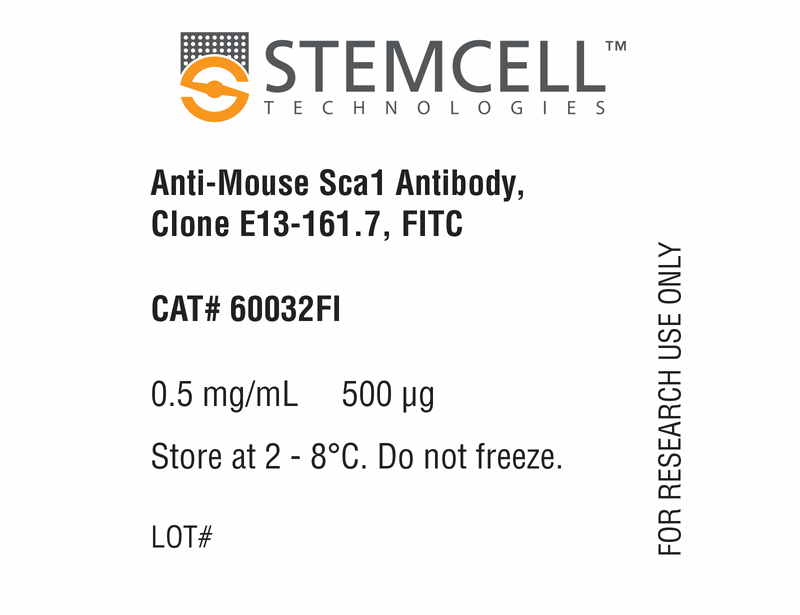
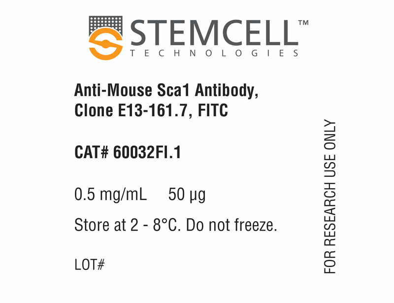
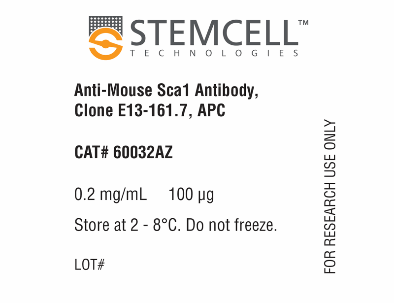
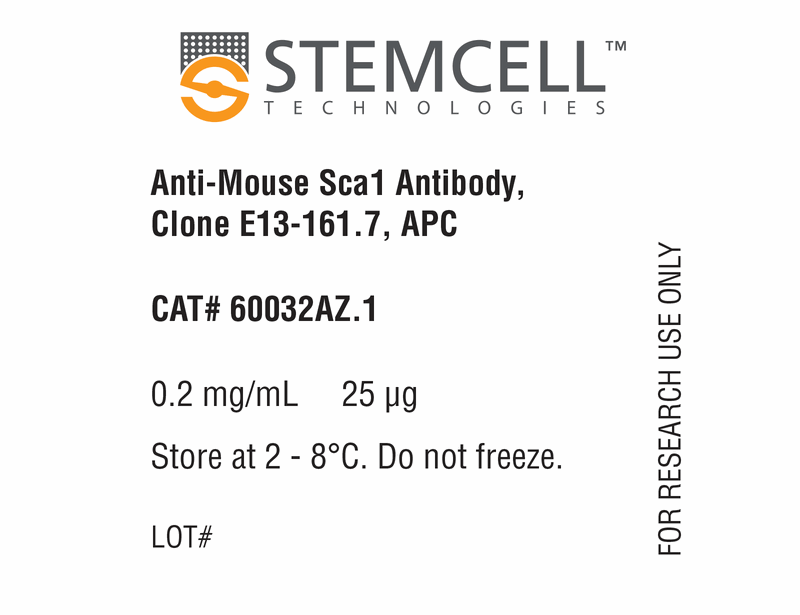
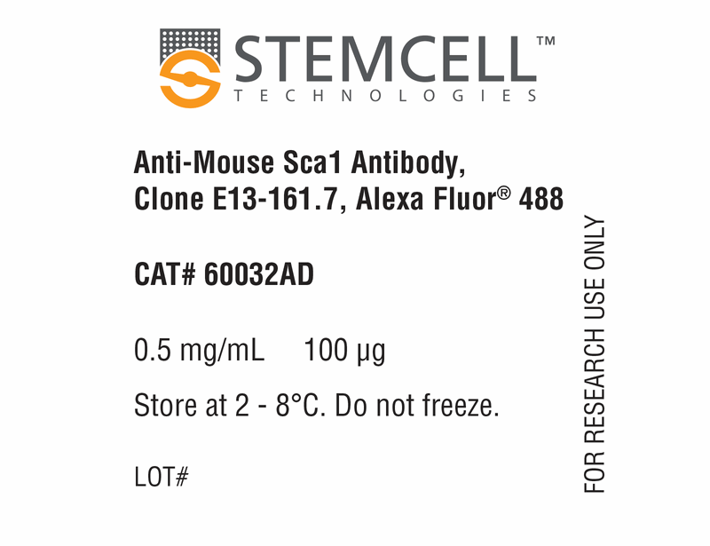
Anti-Mouse Sca1 Antibody, Clone E13-161.7
This antibody clone has been verified for purity assessments of cells isolated with EasySep™ kits, including EasySep™ Mouse SCA1 Biotin Positive Selection Kit (Catalog #18856).
Data
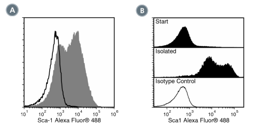
Figure 1. Data for Alexa Fluor® 488-Conjugated
(A) Flow cytometry analysis of C57BL/6 mouse splenocytes labeled with Anti-Mouse Sca1 Antibody, Clone E13-161.7, Alexa Fluor® 488 (filled histogram) or a rat IgG2a, kappa Alexa Fluor® 488 isotype control antibody (open histogram).
(B) Flow cytometry analysis of C57BL/6 mouse bone marrow cells pre-labeled with Anti-Mouse Sca1 Antibody, Clone E13-161.7, Alexa Fluor® 488 and processed with the EasySep™ Mouse SCA1 Positive Selection Kit. Histograms show labeling of bone marrow (Start) and isolated cells (Isolated). Labeling of start cells with a rat IgG2a, kappa Alexa Fluor® 488 isotype control antibody is shown in the bottom panel (open histogram).
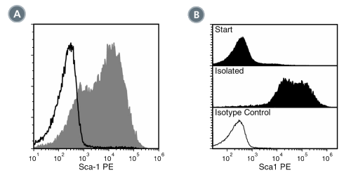
Figure 2. Data for PE-Conjugated
(A) Flow cytometry analysis of C57BL/6 mouse splenocytes labeled with Anti-Mouse Sca1 Antibody, Clone E13-161.7, PE (filled histogram) or a rat IgG2a, kappa PE isotype control antibody (open histogram).
(B) Flow cytometry analysis of C57BL/6 mouse bone marrow cells processed with the EasySep™ PE Selection Kit for Mouse Cells (Catalog #18554) using Anti-Mouse Sca1 Antibody, Clone E13-161.7, PE. Histograms show labeling of bone marrow (Start) and isolated cells (Isolated). Labeling of start cells with a rat IgG2a, kappa PE isotype control antibody is shown in the bottom panel (open histogram).
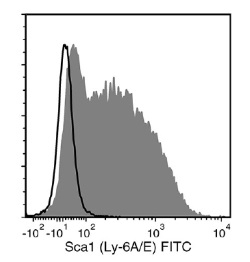
Figure 3. Data for Unconjugated
Flow cytometry analysis of C57BL/6 mouse splenocytes labeled with Anti-Mouse Sca1 Antibody, Clone E13-161.7, followed by a mouse anti-rat IgG2a antibody, FITC (filled histogram), or Rat IgG2a, kappa Isotype Control Antibody, Clone RTK2758 (Catalog #60076), followed by a mouse anti-rat IgG2a antibody, FITC (solid line histogram).
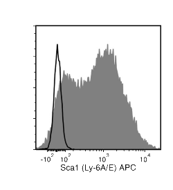
Figure 4. Data for APC-Conjugated
Flow cytometry analysis of C57BL/6 mouse splenocytes labeled with Anti-Mouse Sca1 Antibody, Clone E13-161.7, APC (filled histogram) or a rat IgG2a, kappa isotype control antibody, APC (solid line histogram).
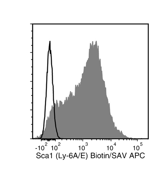
Figure 5. Data for Biotin-Conjugated
Flow cytometry analysis of C57BL/6 mouse splenocytes labeled with Anti-Mouse Sca1 Antibody, Clone E13-161.7, Biotin followed by streptavidin (SAV) APC (filled histogram) or a biotinylated rat IgG2a, kappa isotype control antibody followed by SAV APC (solid line histogram).
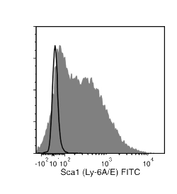
Figure 6. Data for FITC-Conjugated
Flow cytometry analysis of C57BL/6 mouse splenocytes labeled with Anti-Mouse Sca1 Antibody, Clone E13-161.7, FITC (filled histogram) or a rat IgG2a, kappa isotype control antibody, FITC (solid line histogram).

