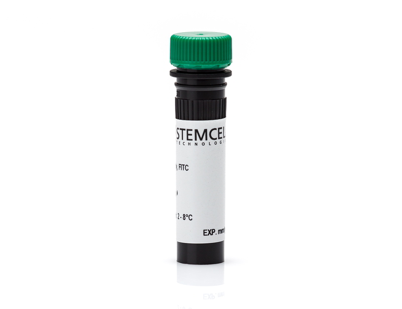
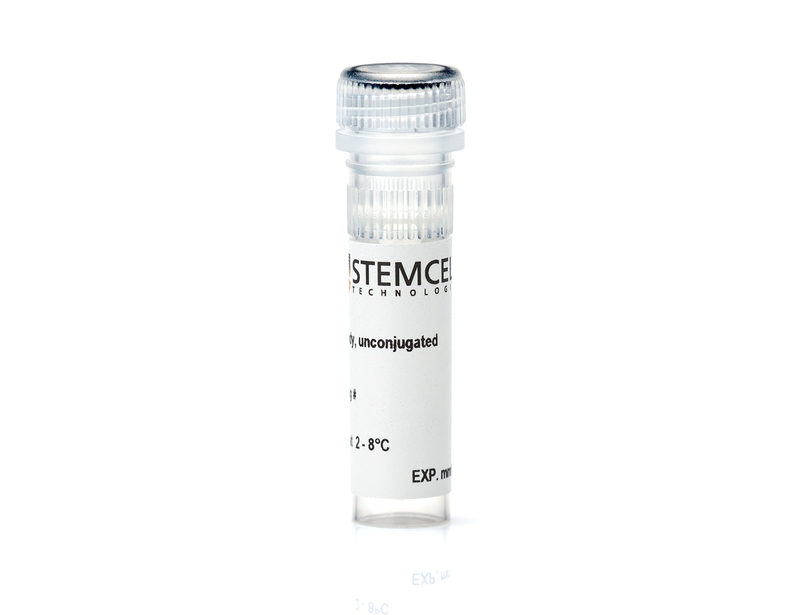
Anti-Mouse EPCR Antibody, Clone RMEPCR1560 (1560)
| Document Type | 产品名称 | Catalog # | Lot # | 语言 |
|---|---|---|---|---|
| Product Information Sheet | Anti-Mouse EPCR Antibody, Clone RMEPCR1560 (1560) | 60038 | All | English |
| Product Information Sheet | Anti-Mouse EPCR Antibody, Clone RMEPCR1560 (1560), Biotin | 60038BT | All | English |
| Product Information Sheet | Anti-Mouse EPCR Antibody, Clone RMEPCR1560 (1560), FITC | 60038FI, 60038FI.1 | All | English |
| Product Information Sheet | Anti-Mouse EPCR Antibody, Clone RMEPCR1560 (1560), PE | 60038PE, 60038PE.1 | All | English |
| Safety Data Sheet | Anti-Mouse EPCR Antibody, Clone RMEPCR1560 (1560) | 60038 | All | English |
| Safety Data Sheet | Anti-Mouse EPCR Antibody, Clone RMEPCR1560 (1560), Biotin | 60038BT | All | English |
| Safety Data Sheet | Anti-Mouse EPCR Antibody, Clone RMEPCR1560 (1560), FITC | 60038FI, 60038FI.1 | All | English |
| Safety Data Sheet | Anti-Mouse EPCR Antibody, Clone RMEPCR1560 (1560), PE | 60038PE, 60038PE.1 | All | English |
Data
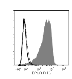
Figure 1. Data for Unconjugated
Flow cytometry analysis of HEK-293 mEPCR-transfected cells (filled histogram) or non-transfected HEK-293 cells (negative control; dashed line histogram), labeled with Anti-Mouse EPCR Antibody, Clone RMEPCR1560, followed by Goat Anti-Mouse IgG (H+L) Antibody, Polyclonal, FITC (Catalog #60138FI). Labeling of HEK-293 mEPCR-transfected cells with a rat IgG2b, kappa isotype control antibody, followed by Goat Anti-Mouse IgG (H+L) Antibody, Polyclonal, FITC is shown (solid line histogram).
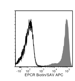
Figure 2. Data for Biotin-Conjugated
Flow cytometry analysis of HEK-293 mEPCR-transfected cells (filled histogram) or non-transfected HEK-293 cells (negative control cells, dashed line histogram), labeled with Anti-Mouse EPCR Antibody, Clone RMEPCR1560, Biotin followed by streptavidin (SAV) APC. Labeling of HEK-293 mEPCR-transfected cells with a rat IgG2b, kappa isotype control antibody followed by SAV APC is shown (solid line histogram).
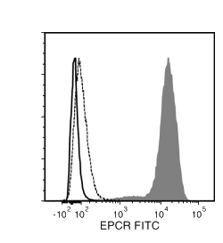
Figure 3. Data for FITC-Conjugated
Flow cytometry analysis of HEK-293 mEPCR-transfected cells (filled histogram) or non-transfected HEK-293 cells (negative control cells, dashed line histogram), labeled with Anti-Mouse EPCR Antibody, Clone RMEPCR1560 FITC. Labeling of HEK-293 mEPCR-transfected cells with a rat IgG2b, kappa FITC isotype control antibody is shown (solid line histogram).
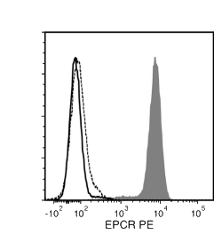
Figure 4. Data for PE-Conjugated
Flow cytometry analysis of HEK-293 mEPCR-transfected cells (filled histogram) or non-transfected HEK-293 cells (negative control cells, dashed line histogram), labeled with Anti-Mouse EPCR Antibody, Clone RMEPCR1560, PE. Labeling of HEK-293 mEPCR-transfected cells with a rat IgG2b, kappa PE isotype control antibody is shown (solid line histogram).


