
Anti-Mouse CD150 Antibody, Clone TC15-12F12.2


Data
Figure 1. Data for Alexa Fluor® 488-Conjugated
Flow cytometry analysis of C57BL/6 mouse splenocytes labeled with Anti-Mouse CD150 Antibody, Clone TC15-12F12.2, Alexa Fluor® 488 (filled histogram) or a rat IgG2a, lambda Alexa Fluor® 488 isotype control antibody (solid line histogram).
Figure 2. Data for PE-Conjugated
(A) Flow cytometry analysis of C57BL/6 mouse splenocytes labeled with Anti-Mouse CD150 Antibody, Clone TC15-12F12.2, PE (filled histogram) or a rat IgG2a, lambda PE isotype control antibody (solid line histogram). (B) Flow cytometry analysis of C57BL/6 mouse bone marrow cells labeled with Anti-Mouse CD150 Antibody, Clone TC15-12F12.2, PE (filled histogram) or a rat IgG2a, lambda PE isotype control antibody (solid line histogram).
Figure 3. Data for Unconjugated
Flow cytometry analysis of C57BL/6 mouse splenocytes labeled with Anti-Mouse CD150 Antibody, Clone TC15-12F12.2, followed by a mouse anti-rat IgG2a antibody, FITC (filled histogram), or a rat IgG2a isotype control antibody followed by a mouse anti-rat IgG2a antibody, FITC (solid line histogram).
Figure 4. Data for APC-Conjugated
Flow cytometry analysis of C57BL/6 mouse splenocytes labeled with Anti-Mouse CD150 Antibody, Clone TC15-12F12.2, APC (filled histogram) or a rat IgG2a isotype control antibody, APC (solid line histogram).
Figure 5. Data for Biotin-Conjugated
Flow cytometry analysis of C57BL/6 mouse splenocytes labeled with Anti-Mouse CD150 Antibody, Clone TC15-12F12.2, Biotin followed by streptavidin (SAV) APC (filled histogram), or a biotinylated rat IgG2a isotype control antibody followed by SAV APC (solid line histogram).
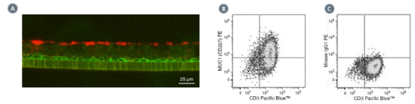
(A) Primary human airway epithelial cells were cultured in PneumaCult™-ALI Medium (Catalog #05001) at the air-liquid interface, then cryo-sectioned and labeled with Anti-Human MUC1 (CD227) Antibody, Clone 16A, followed by a goat anti-rabbit IgG antibody, Alexa Fluor® 594 (red), and an anti-human NGF Receptor/p75NTR (CD271) antibody, followed by a donkey anti-mouse IgG antibody, Alexa Fluor® 488 (green). (B) Flow cytometry analysis of human peripheral blood lymphocytes following stimulation with PHA for 3 days. Cells were labeled with Anti-Human MUC1 (CD227) Antibody, Clone 16A, followed by an anti-mouse IgG1 antibody, PE and anti-human CD3 antibody, Clone HIT3a, Pacific Blue™. (C) Flow cytometry analysis of human peripheral blood lymphocytes following stimulation with PHA for 3 days. Cells were labeled with Mouse IgG1, kappa Isotype Control Antibody, Clone MOPC-21 (Catalog #60070), followed by an anti-mouse IgG1 antibody, PE, and anti-human CD3 antibody, clone HIT3a, Pacific Blue™.

(A) Flow cytometry analysis of human airway epithelial cells cultured in PneumaCult™-ALI Medium (Catalog #05001) at the air-liquid interface. Cells were enzymatically dissociated and labeled with Anti-Human MUC1 (CD227) Antibody, Clone 16A, PE (filled histogram) or Mouse IgG1, kappa Isotype Control Antibody, Clone MOPC-21, PE (Catalog #60070PE, solid line histogram). (C) Flow cytometry analysis human peripheral blood lymphocytes following stimulation with PHA for 3 days. Cells were labeled with Anti-Human MUC1 (CD227) Antibody, Clone 16A, PE and anti-human CD3 antibody, clone HIT3a, Pacific Blue™. (D) Flow cytometry analysis of PHA-activated human peripheral blood lymphocytes following stimulation with PHA for 3 days. Cells were labeled with Mouse IgG1, kappa Isotype Control Antibody, Clone MOPC-21, PE, and anti-human CD3 antibody, clone HIT3a, Pacific Blue™.
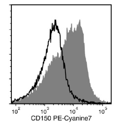
Flow cytometry analysis of C57BL/6 mouse splenocytes labeled with Anti-Mouse CD150 Antibody, Clone TC15-12F12.2, PE-Cyanine7 (filled histogram) or a rat IgG2a isotype control antibody, PE-Cyanine7 (solid line histogram).