Anti-Mouse CD44 Antibody, Clone IM7

Rat monoclonal IgG2b antibody against human, mouse, rhesus CD44 (tissue non-specific alkaline phosphatase)
概要
The IM7 antibody reacts with CD44 (Ly-24), an ~80 - 95 kDa type 1 transmembrane glycoprotein involved in cell-cell and cell-matrix interactions. CD44 is expressed on the surface of many cells, including leukocytes and hepatocytes, as well as endothelial, epithelial, and mesenchymal cells. Expression levels increase upon activation of T and B cells, and memory cells exhibit a CD44high phenotype. CD44 binds many ligands, including hyaluronic acid, collagen, fibronectin, growth factors, and metalloproteinases, thus modulating processes such as lymphocyte activation, recirculation and homing, leukocyte rolling and aggregation, hematopoiesis, and tumor metastasis. Numerous disorders are associated with altered expression or dysfunction of CD44. Many CD44 isoforms have been identified, with alternative splicing, differential N- and O- glycosylation, and sulfation mediating the functional role(s) played by the protein in a specific cell. The IM7 monoclonal antibody reacts with an extracellular epitope found on all isoforms of CD44 and both murine allotypes.

This antibody clone has been verified for purity assessments of cells isolated with EasySep™ kits, including EasySep™ Mouse CD4+ T Cell Isolation Kit (Catalog #19852), EasySep™ Mouse CD4+CD62L+ T Cell Isolation Kit (Catalog #18765), and EasySep™ Human Naïve CD4+ T Cell Isolation Kit (Catalog #19555).
Subtype
Primary Antibodies
Target Antigen
CD44
Alternative Names
ECMR III, gp85, H-CAM, Hermes, HUTCH-1, Ly24, Ly-24, Pgp-1
Reactive Species
Baboon, Cynomolgus, Rhesus, Squirrel Monkey, Cat, Chimpanzee, Cow, Dog, Horse, Human, Mouse, Pig
Conjugation
Alexa Fluor 488, APC, Biotin, FITC, PE, PerCP-Cyanine5.5, Unconjugated
Host Species
Rat
Cell Type
Mammary Cells, T Cells
Species
Human, Mouse, Non-Human Primate, Other
Application
Cell Isolation, CyTOF, ELISA, Flow Cytometry, Functional Assay, Immunocytochemistry, Immunofluorescence, Immunohistochemistry, Immunoprecipitation
Area of Interest
Epithelial Cell Biology, Immunology, Stem Cell Biology
Clone
IM7
Gene ID
12505/960
Isotype
IgG2b, kappa
数据及文献

Data

Data for Unconjugated

Figure 1. Data for Unconjugated

Flow cytometry analysis of human peripheral blood mononuclear cells (PBMCs; gated on lymphocytes) labeled with Anti-Mouse CD44 Antibody, Clone IM7, followed by Goat Anti-Mouse IgG (H+L) Antibody, Polyclonal, FITC (Catalog #60138FI) (filled histogram), or a rat IgG2b, kappa isotype control antibody, followed by Goat Anti-Mouse IgG (H+L) Antibody, Polyclonal, FITC (solid line histogram).

Data for Alexa Fluor® 488-Conjugated

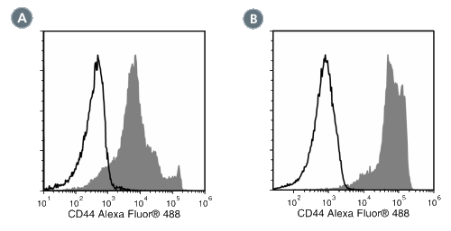
Figure 2. Data for Alexa Fluor® 488-Conjugated

(A) Flow cytometry analysis of C57BL/6 mouse splenocytes labeled with Anti-Mouse CD44 Antibody, Clone IM7, Alexa Fluor® 488 (filled histogram) or a rat IgG2b, kappa Alexa Fluor® 488 isotype control antibody (open histogram).
(B) Flow cytometry analysis of human peripheral blood mononuclear cells (PBMCs) labeled with Anti-Mouse CD44 Antibody, Clone IM7, Alexa Fluor® 488 (filled histogram) or a rat IgG2b, kappa Alexa Fluor® 488 isotype control antibody (open histogram).

Data for Biotin-Conjugated

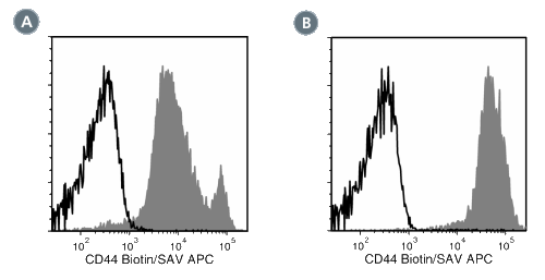
Figure 3. Data for Biotin-Conjugated

(A) Flow cytometry analysis of C57BL/6 mouse splenocytes labeled with Anti-Mouse CD44 Antibody, Clone IM7, Biotin followed by streptavidin (SAV) APC (filled histogram) or a rat IgG2b, kappa Biotin isotype control antibody followed by SAV APC (solid line histogram).
(B) Flow cytometry analysis of human peripheral blood mononuclear cells (PBMCs) labeled with Anti-Mouse CD44 Antibody, Clone IM7, Biotin followed by streptavidin (SAV) APC (filled histogram) or a rat IgG2b, kappa Biotin isotype control antibodyfollowed by SAV APC (open histogram).

Data for PE-Conjugated

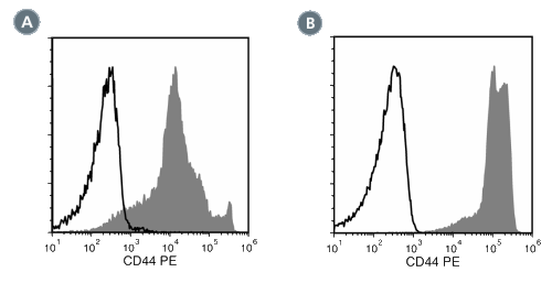
Figure 4. Data for PE-Conjugated

(A) Flow cytometry analysis of C57BL/6 mouse splenocytes labeled with Anti-Mouse CD44 Antibody, Clone IM7, PE (filled histogram) or a rat IgG2b, kappa PE isotype control antibody (open histogram).
(B) Flow cytometry analysis of human peripheral blood mononuclear cells (PBMCs) labeled with Anti-Mouse CD44 Antibody, Clone IM7, PE (filled histogram) or a rat IgG2b, kappa PE isotype control antibody (open histogram).

Data for PerCP-Cy55-Conjugated

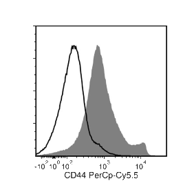
Figure 5. Data for PerCP-Cy55-Conjugated

Flow cytometry analysis of C57BL/6 mouse splenocytes labeled with Anti-Mouse CD44 Antibody, Clone IM7, PerCP-Cy5.5 (filled histogram) or a rat IgG2b, kappa isotype control antibody, PerCP-Cy5.5 (solid line histogram).

Data for FITC-Conjugated

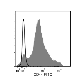
Figure 6. Data for FITC-Conjugated

Flow cytometry analysis of C57BL/6 mouse splenocytes labeled with Anti-Mouse CD44 Antibody, Clone IM7, FITC (filled histogram) or Rat IgG2b, kappa Isotype Control Antibody, Clone RTK4530, FITC (Catalog #60077FI) (solid line histogram).

Data for APC-Conjugated

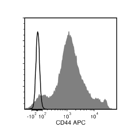
Figure 7. Data for APC-Conjugated

Flow cytometry analysis of C57BL/6 mouse splenocytes labeled with Anti-Mouse CD44 Antibody, Clone IM7, APC (filled histogram) or Rat IgG2b, kappa Isotype Control Antibody, Clone RTK4530, APC (Catalog #60077AZ) (solid line histogram).

Publications (1)

Cell 2018 JAN Transmembrane Pickets Connect Cyto- and Pericellular Skeletons Forming Barriers to Receptor Engagement. Freeman SA et al.

Abstract

Phagocytic receptors must diffuse laterally to become activated upon clustering by multivalent targets. Receptor diffusion, however, can be obstructed by transmembrane proteins (pickets") that are immobilized by interacting with the cortical cytoskeleton. The molecular identity of these pickets and their role in phagocytosis have not been defined. We used single-molecule tracking to study the interaction between Fcγ receptors and CD44 an abundant transmembrane protein capable of indirect association with F-actin hence likely to serve as a picket. CD44 tethers reversibly to formin-induced actin filaments curtailing receptor diffusion. Such linear filaments predominate in the trailing end of polarized macrophages where receptor mobility was minimal. Conversely receptors were most mobile at the leading edge where Arp2/3-driven actin branching predominates. CD44 binds hyaluronan anchoring a pericellular coat that also limits receptor displacement and obstructs access to phagocytic targets. Force must be applied to traverse the pericellular barrier enabling receptors to engage their targets.
View All Publications
Top