
Anti-Human CD105 Antibody, Clone 43A3


This antibody clone has been verified for labeling human mesenchymal cells grown in MesenCult™ Proliferation Kit (Human; Catalog #05411) and MesenCult™-XF Medium (Catalog #05420).
Data
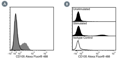
Figure 1. Data for Alexa Fluor® 488-Conjugated
(A) Flow cytometry analysis of human peripheral blood mononuclear cells (PBMCs) labeled with Anti-Human CD105 Antibody, Clone 43A3, Alexa Fluor® 488 (filled histogram) or a mouse IgG1, kappa Alexa Fluor® 488 isotype control antibody (solid line histogram). (B) Flow cytometry analysis of human PBMCs cultured for 24 hours with or without lipopolysaccharide (LPS) and labeled with Anti-Human CD105 Antibody, Clone 43A3, Alexa Fluor® 488. Histograms show labeling of PBMCs cultured in the absence (Unstimulated) or presence (Stimulated) of LPS. Labeling of LPS-stimulated PBMCs with a mouse IgG1, kappa isotype control antibody, Alexa Fluor® 488 is shown (solid line histogram).
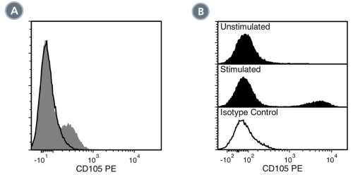
Figure 2. Data for PE-Conjugated
(A) Flow cytometry analysis of human peripheral blood mononuclear cells (PBMCs) labeled with Anti-Human CD105 Antibody, Clone 43A3, PE (filled histogram) or a mouse IgG1, kappa PE isotype control antibody (solid line histogram). (B) Flow cytometry analysis of human PBMCs cultured for 24 hours with or without lipopolysaccharide (LPS) and labeled with Anti-Human CD105 Antibody, Clone 43A3, PE. Histograms show labeling of PBMCs cultured in the absence (Unstimulated) or presence (Stimulated) of LPS. Labeling of LPS-stimulated PBMCs with a mouse IgG1, kappa isotype control antibody, PE is shown (solid line histogram).
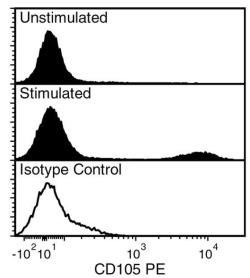
Figure 3. Data for Unconjugated
Flow cytometry analysis of human peripheral blood mononuclear cells (PBMCs) cultured for 24 hours with or without lipopolysaccharide (LPS) and labeled with Anti-Human CD105 Antibody, Clone 43A3, followed by a rat anti-mouse IgG1 antibody, PE. Histograms show labeling of PBMCs cultured in the absence (Unstimulated) or presence (Stimulated) of LPS. Labeling of LPS-stimulated PBMCs with a mouse IgG1, kappa isotype control antibody, followed by a rat anti-mouse IgG1 antibody, PE is shown (solid line histogram).
Figure 4. Data for APC-Conjugated
Flow cytometry analysis of human peripheral blood mononuclear cells (PBMCs) cultured for 24 h with or without lipopolysaccharide (LPS) and labeled with Anti-Human CD105 Antibody, Clone 43A3, APC. Histograms show labeling of PBMCs cultured in the absence (Unstimulated) or presence (Stimulated) of LPS. Labeling of LPS-stimulated PBMCs with a mouse IgG1, kappa isotype control antibody, APC is shown in the bottom panel (solid line histogram).
Figure 5. Data for Biotin-Conjugated
Flow cytometry analysis of human peripheral blood mononuclear cells (PBMCs) cultured for 24 h with or without lipopolysaccharide (LPS) and labeled with Anti-Human CD105 Antibody, Clone 43A3, Biotin followed by streptavidin (SAV) APC. Histograms show labeling of PBMCs cultured in the absence (Unstimulated) or presence (Stimulated) of LPS. Labeling of LPS-stimulated PBMCs with a biotinylated mouse IgG1, kappa isotype control antibody followed by SAV APC is shown in the bottom panel (solid line histogram).
Figure 6. Data for FITC-Conjugated
Flow cytometry analysis of human peripheral blood mononuclear cells (PBMCs) cultured for 24 h with or without lipopolysaccharide (LPS) and labeled with Anti-Human CD105 Antibody, Clone 43A3, FITC. Histograms show labeling of PBMCs cultured in the absence (Unstimulated) or presence (Stimulated) of LPS. Labeling of LPS-stimulated PBMCs with a mouse IgG1, kappa isotype control antibody, FITC is shown in the bottom panel (solid line histogram).