
Anti-Human CD81 (TAPA-1) Antibody, Clone 5A6

CD81, a 26 kDa non-glycosylated type III transmembrane protein, belongs to the tetraspanin family. Tetraspanins contain four transmembrane domains, two extracellular loops, and short cytoplasmic N- and C-termini. CD81 associates with numerous integrins, co-receptors, and other proteins to form multimolecular complexes in the plasma membrane called tetraspanin-enriched microdomains. Palmitoylation of the juxtamembrane cysteine residues is important for its association with other proteins and the plasma membrane.
CD81 modulates a diverse array of biological functions including cell-cell adhesion and fusion, cell migration, and proliferation. For example, together with CD19 and CD21, CD81 forms a co-receptor for the B cell receptor on B cells. The epitope of the 5A6 antibody is hidden when CD81 is in complex with CD19 but accessible following activation of the B cell. Mutations in CD81 have been associated with the immune disorder common variable immunodeficiency (CVID). CD81 has also been implicated in mediating the entry of hepatitis C virus into cells.
| Document Type | 产品名称 | Catalog # | Lot # | 语言 |
|---|---|---|---|---|
| Product Information Sheet | Anti-Human CD81 (TAPA-1) Antibody, Clone 5A6 | 100-0209 | All | English |
| Special Protocol | Anti-Human CD81 (TAPA-1) Antibody, Clone 5A6 | 100-0209 | All | English |
| Safety Data Sheet | Anti-Human CD81 (TAPA-1) Antibody, Clone 5A6 | 100-0209 | All | English |
Data
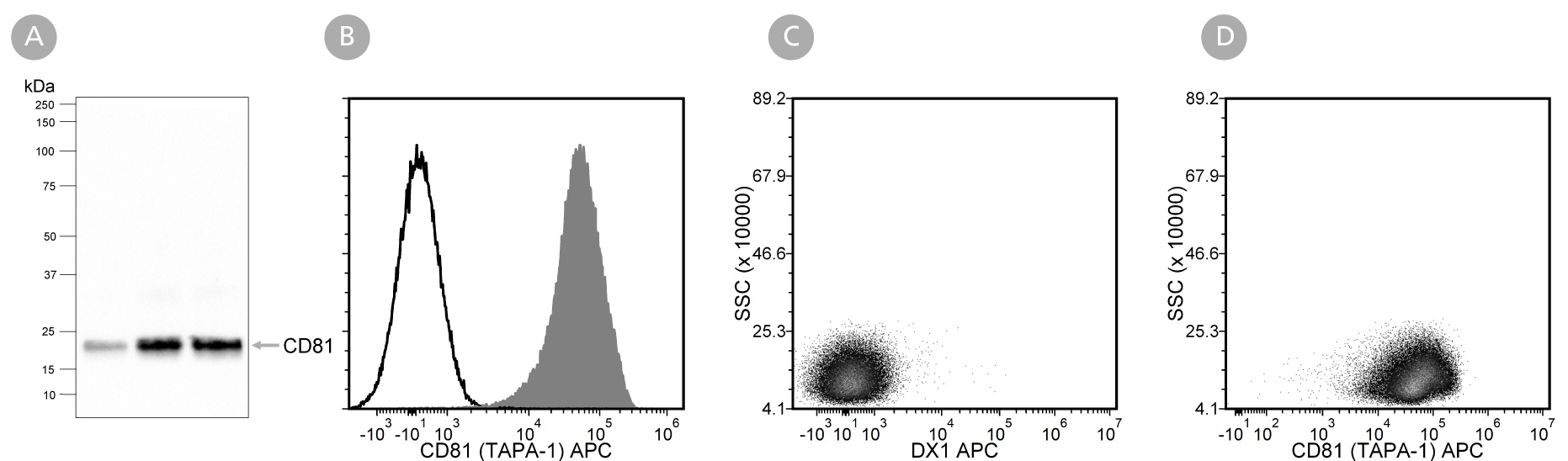
Figure 1. Analysis of Anti-Human CD81 (TAPA-1) Antibody, Clone 5A6 with Western Blot and Flow Cytometry
Extracellular vesicles (EVs) isolated from mesenchymal stromal cell (MSC)-conditioned medium using a 2 mL EV size exclusion chromatography column were analysed with (A) western blot analysis with Anti-Human CD81 (TAPA-1) Antibody, Clone 5A6. Lanes (left to right) were loaded with isolated fractions 9, 10, and 11, respectively. Further validation was performed by flow cytometry analysis of human peripheral blood mononuclear cells (PBMCs; viable lymphocytes were gated for analysis) stimulated with phorbol myristate acetate (PMA) and ionomycin and then (B) labeled with Anti-Human CD81 (TAPA-1) Antibody, Clone 5A6, followed by a goat anti-mouse IgG1 antibody, APC (filled histogram), or Anti-Dextran Antibody, Clone DX1 (Catalog #60026) as an isotype control, followed by a goat anti-mouse IgG1 antibody, APC (solid line histogram), or (C) labeled with Anti-Dextran Antibody, Clone DX1 (Catalog #60026) as an isotype control, followed by a goat anti-mouse IgG1 antibody, APC, or (D) labeled with Anti-Human CD81 (TAPA-1) Antibody, Clone 5A6, followed by a goat anti-mouse IgG1 antibody, APC.