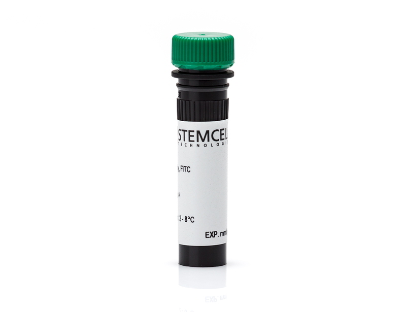
Anti-Human CD66b Antibody, Clone G10F5
| Document Type | 产品名称 | Catalog # | Lot # | 语言 |
|---|---|---|---|---|
| Product Information Sheet | Anti-Human CD66b Antibody, Clone G10F5 | 60086 | All | English |
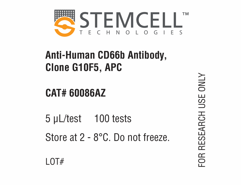
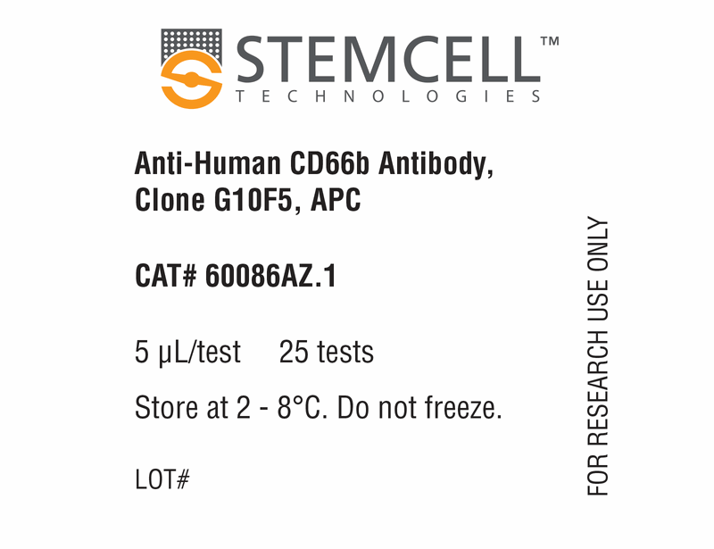
| Product Information Sheet | Anti-Human CD66b Antibody, Clone G10F5, APC | 60086AZ, 60086AZ.1 | All | English |
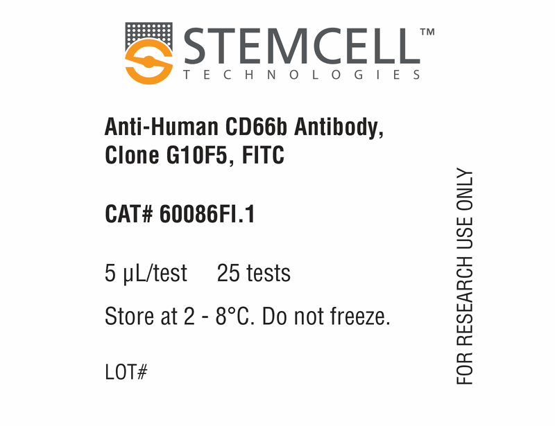
| Product Information Sheet | Anti-Human CD66b Antibody, Clone G10F5, FITC | 60086FI, 60086FI.1 | All | English |
| Product Information Sheet | Anti-Human CD66b Antibody, Clone G10F5, PE | 60086PE, 60086PE.1 | All | English |
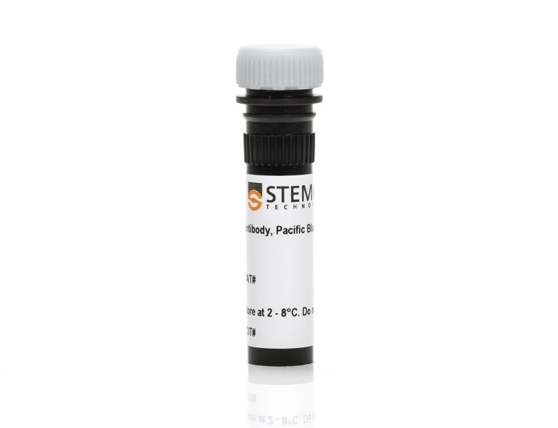
| Product Information Sheet | Anti-Human CD66b Antibody, Clone G10F5, Pacific Blue™ | 60086PB, 60086PB.1 | All | English |
| Safety Data Sheet | Anti-Human CD66b Antibody, Clone G10F5 | 60086 | All | English |
| Safety Data Sheet | Anti-Human CD66b Antibody, Clone G10F5, APC | 60086AZ, 60086AZ.1 | All | English |
| Safety Data Sheet | Anti-Human CD66b Antibody, Clone G10F5, FITC | 60086FI, 60086FI.1 | All | English |
| Safety Data Sheet | Anti-Human CD66b Antibody, Clone G10F5, PE | 60086PE, 60086PE.1 | All | English |
| Safety Data Sheet | Anti-Human CD66b Antibody, Clone G10F5, Pacific Blue™ | 60086PB, 60086PB.1 | All | English |
Data
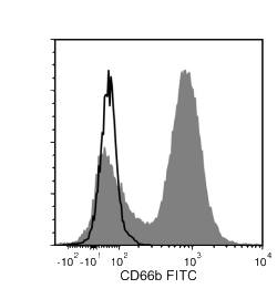
Figure 1. Data for Unconjugated
Flow cytometry analysis of human whole blood nucleated cells labeled with Anti-Human CD66b Antibody, Clone G10F5, followed by Goat Anti-Mouse IgG (H+L) Antibody, Polyclonal, FITC (Catalog #60138FI) (filled histogram), or Mouse IgM, kappa Isotype Control Antibody, Clone MM-30 (Catalog #60069), followed by Goat Anti-Mouse IgG (H+L) Antibody, Polyclonal, FITC (solid line histogram).
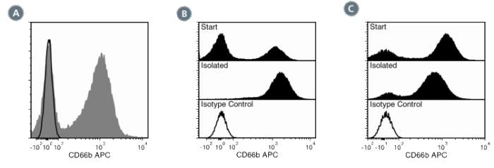
Figure 2. Data for APC-Conjugated
(A) Flow cytometry analysis of human whole blood nucleated cells labeled with Anti-Human CD66b Antibody, Clone G10F5, APC (filled histogram) or a mouse IgM, kappa APC isotype control antibody (solid line histogram). (B) Flow cytometry analysis of human buffy coat nucleated cells processed with the EasySep™ HLA Whole Blood CD15 Positive Selection Kit and labeled with Anti-Human CD66b Antibody, Clone G10F5, APC. Histograms show labeling of buffy coat nucleated cells (Start) and isolated cells (Isolated). Labeling of start cells with a mouse IgM, kappa APC isotype control antibody is shown (solid line histogram). (C) Flow cytometry analysis of whole blood nucleated cells processed with the EasySep™ Human Myeloid Positive Selection Kit and labeled with Anti-Human CD66b Antibody, Clone G10F5, APC. Histograms show labeling of whole blood nucleated cells (Start) and isolated cells (Isolated). Labeling of start cells with a mouse IgM, kappa APC isotype control antibody is shown (solid line histogram).
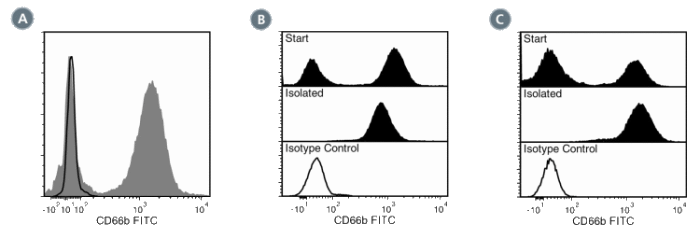
Figure 3. Data for FITC-Conjugated
(A) Flow cytometry analysis of human whole blood nucleated cells labeled with Anti-Human CD66b Antibody, Clone G10F5, FITC (filled histogram) or a mouse IgM, kappa FITC isotype control antibody (solid line histogram). (B) Flow cytometry analysis of human whole blood nucleated cells processed with the EasySep™ Whole Blood CD66b Positive Selection Kit and labeled with Anti-Human CD66b Antibody, Clone G10F5, FITC. Histograms show labeling of whole blood nucleated cells (Start) and isolated cells (Isolated). Labeling of start cells with a mouse IgM, kappa FITC isotype control antibody is shown (solid line histogram). (C) Flow cytometry analysis of human buffy coat nucleated cells processed with the EasySep™ HLA Whole Blood CD15 Positive Selection Kit and labeled with Anti-Human CD66b Antibody, Clone G10F5, FITC. Histograms show labeling of buffy coat nucleated cells (Start) and isolated cells (Isolated). Labeling of start cells with a mouse IgM, kappa FITC isotype control antibody is shown (solid line histogram).
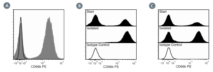
Figure 4. Data for PE-Conjugated
(A) Flow cytometry analysis of human whole blood nucleated cells labeled with Anti-Human CD66b Antibody, Clone G10F5, PE (filled histogram) or Mouse IgM, kappa Isotype Control Antibody, Clone MM-30, PE (Catalog #60069PE) (solid line histogram). (B) Flow cytometry analysis of human buffy coat nucleated cells processed with the EasySep™ HLA Whole Blood CD15 Positive Selection Kit and labeled with Anti-Human CD66b Antibody, Clone G10F5, PE. Histograms show labeling of buffy coat nucleated cells (Start) and isolated cells (Isolated). Labeling of start cells with Mouse IgM, kappa Isotype Control Antibody, Clone MM30, PE is shown (solid line histogram). (C) Flow cytometry analysis of whole blood nucleated cells processed with the EasySep™ HLA Whole Blood CD33 Positive Selection Kit and labeled with AntiHuman CD66b Antibody, Clone G10F5, PE. Histograms show labeling of whole blood nucleated cells (Start) and isolated cells (Isolated). Labeling of start cells with Mouse IgM, kappa Isotype Control Antibody, Clone MM-30, PE is shown (solid line histogram).

Figure 5. Data for PB-Conjugated
Flow cytometry analysis of human peripheral blood mononuclear cells (PBMCs) labeled with Anti-Human CD66b Antibody, Clone G10F5, Pacific Blue™ (filled histogram) or a mouse IgM, kappa Pacific Blue™ isotype control antibody (solid line histogram).

