
Anti-Human CD51 Antibody, Clone NKI-M9

| Document Type | 产品名称 | Catalog # | Lot # | 语言 |
|---|---|---|---|---|
| Product Information Sheet | Anti-Human CD51 Antibody, Clone NKI-M9 | 60043 | All | English |
| Product Information Sheet | Anti-Human CD51 Antibody, Clone NKI-M9, Biotin | 60043BT | All | English |
| Product Information Sheet | Anti-Human CD51 Antibody, Clone NKI-M9, FITC | 60043FI, 60043FI.1 | All | English |
| Product Information Sheet | Anti-Human CD51 Antibody, Clone NKI-M9, PE | 60043PE, 60043PE.1 | All | English |
| Safety Data Sheet | Anti-Human CD51 Antibody, Clone NKI-M9 | 60043 | All | English |
| Safety Data Sheet | Anti-Human CD51 Antibody, Clone NKI-M9, Biotin | 60043BT | All | English |
| Safety Data Sheet | Anti-Human CD51 Antibody, Clone NKI-M9, FITC | 60043FI, 60043FI.1 | All | English |
| Safety Data Sheet | Anti-Human CD51 Antibody, Clone NKI-M9, PE | 60043PE, 60043PE.1 | All | English |
Data
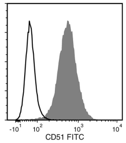
Figure 1. Data for Unconjugated
Flow cytometry analysis of HT1080 human fibrosarcoma cells labeled with Anti-Human CD51 Antibody, Clone NKI-M9, followed by Goat Anti-Mouse IgG (H+L) Antibody, Polyclonal, FITC (Catalog #60138FI) (filled histogram), or Mouse IgG2a, kappa Isotype Control Antibody, Clone MOPC173 (Catalog #60071), followed by Goat Anti-Mouse IgG (H+L) Antibody, Polyclonal, FITC (solid line histogram).
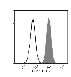
Figure 2. Data for FITC-Conjugated
Flow cytometry analysis of human HT-1080 fibrosarcoma cells labeled with Anti-Human CD51 Antibody, Clone NKI-M9, FITC (filled historgram) or a mouse IgG2a, kappa FITC isotype control antibody (open histogram).
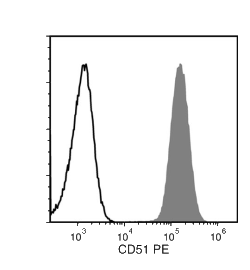
Figure 3. Data for PE-Conjugated
Flow cytometry analysis of human HT1080 fibrosarcoma cells labeled with Anti-Human CD51 Antibody, Clone NKI-M9, PE (filled histogram) or a mouse IgG2a, kappa PE isotype control antibody (solid line histogram).
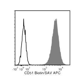
Figure 4. Data for Biotin-Conjugated
Flow cytometry analysis of human HT1080 fibrosarcoma cells labeled with Anti-Human CD51 Antibody, Clone NKI-M9, Biotin, followed by streptavidin (SAV) APC (filled histogram), or Mouse IgG2a, kappa Isotype Control Antibody, Clone MOPC-173, Biotin (Catalog #60071BT), followed by SAV APC (solid line histogram).