Anti-Human CD16 Antibody, Clone 3G8

Mouse monoclonal IgG1 antibody against human, rhesus, cynomolgus CD16
概要
The 3G8 antibody reacts with CD16 (FcγRIII or low affinity IgG receptor III), a type 1 transmembrane glycoprotein belonging to the Ig superfamily. CD16 exists in two distinct isoforms: CD16a, a 50 - 65 kDa form expressed on NK cells, activated monocytes, macrophages, and placental trophoblasts, and CD16b, an ~48 kDa glycosylphosphatidylinositol (GPI)-anchored form expressed on neutrophils, basophils and eosinophils and found as at least two polymorphic variants, termed NA1 and NA2. CD16 binds weakly to the Fc region of monomeric, aggregated or complexed IgG, particularly the IgG1 and IgG3 isotypes. Binding of IgG to either CD16 isoform induces signaling pathways that modulate several types of responses, including antibody-dependent cell-mediated cytotoxicity (ADCC), phagocytosis, cytokine release and proliferation. CD16/IgG interactions can result in non-specific labeling in antibody-based detection and cell separation experiments and the 3G8 antibody may be employed as a blocking antibody to reduce non-specific binding.
Subtype
Primary Antibodies
Target Antigen
CD16
Alternative Names
CD16A, CD16B, Fc-gamma RIII; FCG3; FCGR3; FCGRIII; FcγRIII; FCR-10, FcRIII, IGFR3, IMD20
Reactive Species
Baboon, Capuchin Monkey, Common Marmoset, Cotton-Topped Tamarin, Cynomolgus, Rhesus, Sooty Mangabey, Squirrel Monkey, Chimpanzee, Human, Macaque
Conjugation
Alexa Fluor 488, APC, Biotin, FITC, PE, Unconjugated
Host Species
Mouse
Cell Type
Granulocytes and Subsets, Monocytes
Species
Human, Non-Human Primate
Application
Cell Isolation, CyTOF, Depletion, ELISA, Flow Cytometry, Functional Assay, Immunocytochemistry, Immunofluorescence, Immunohistochemistry, Immunoprecipitation, Neutralization and Blocking
Area of Interest
Immunology
Clone
3G8
Gene ID
2214
Isotype
IgG1, kappa
数据及文献

Data

Data for Unconjugated

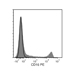
Figure 1. Data for Unconjugated

Flow cytometry analysis of human peripheral blood mononuclear cells (PBMCs) labeled with Anti-Human CD16 Antibody, Clone 3G8, followed by a rat anti-mouse IgG1 antibody, PE (filled histogram), or Mouse IgG1, kappa Isotype Control Antibody, Clone MOPC-21 (Catalog #60070), followed by a rat anti-mouse IgG1 antibody, PE (solid line histogram).

Data for Alexa Fluor® 488-Conjugated

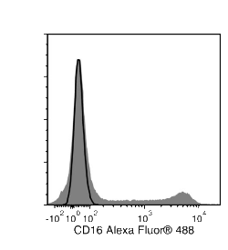
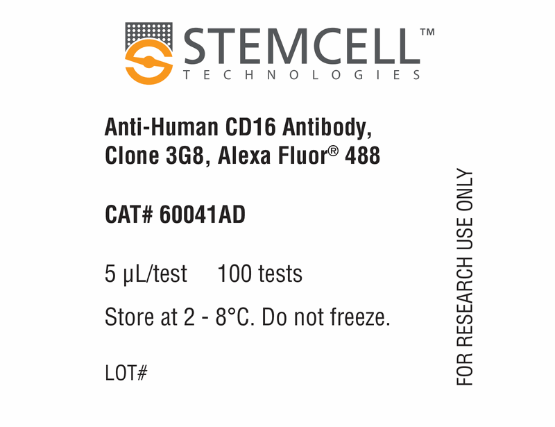
Figure 2. Data for Alexa Fluor® 488-Conjugated

Flow cytometry analysis of human peripheral blood mononuclear cells (PBMCs) labeled with Anti-Human CD16 Antibody, Clone 3G8, Alexa Fluor® 488 (filled histogram) or Mouse IgG1, kappa Isotype Control Antibody, Clone MOPC-21, Alexa Fluor® 488 (Catalog #60070AD) (solid line histogram).

Data for APC-Conjugated

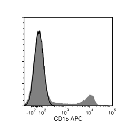
Figure 3. Data for APC-Conjugated

Flow cytometry analysis of human peripheral blood mononuclear cells (PBMCs) labeled with Anti-Human CD16 Antibody, Clone 3G8, APC (filled histogram) or Mouse IgG1, kappa Isotype Control Antibody, Clone MOPC-21, APC (Catalog #60070AZ) (solid line histogram).

Data for Biotin-Conjugated

Figure 4. Data for Biotin-Conjugated

Flow cytometry analysis of human peripheral blood mononuclear cells (PBMCs) labeled with Anti-Human CD16 Antibody, Clone 3G8, Biotin, followed by streptavidin (SAV) APC (filled histogram), or Mouse IgG1, kappa Isotype Control Antibody, Clone MOPC-21, Biotin (Catalog #60070BT), followed by SAV APC (solid line histogram).

Data for FITC-Conjugated

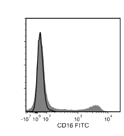
Figure 5. Data for FITC-Conjugated

Flow cytometry analysis of human peripheral blood mononuclear cells (PBMCs) labeled with Anti-Human CD16 Antibody, Clone 3G8, FITC (filled histogram) or Mouse IgG1, kappa Isotype Control Antibody, Clone MOPC-21, FITC (Catalog #60070FI) (solid line histogram).

Data for PE-Conjugated

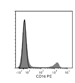
Figure 6. Data for PE-Conjugated

Flow cytometry analysis of human peripheral blood mononuclear cells (PBMCs) labeled with Anti-Human CD16 Antibody, Clone 3G8, PE (filled histogram) or Mouse IgG1, kappa Isotype Control Antibody, Clone MOPC-21, PE (Catalog #60070PE) (solid line histogram).

Top