
Anti-Human CD9 Antibody, Clone HI9a
CD9 belongs to the tetraspanin family of proteins, which contain four transmembrane domains, two extracellular loops, and short cytoplasmic N- and C-termini. It is involved in mediating several cellular processes, including angiogenesis, by promoting cell adhesion, migration, and signal transduction, and is involved in cell fusion processes linked to fertilization, osteoclastogenesis, and myogenesis.
| Document Type | 产品名称 | Catalog # | Lot # | 语言 |
|---|---|---|---|---|
| Product Information Sheet | Anti-Human CD9 Antibody, Clone HI9a | 100-0138 | All | English |
| Special Protocol | Anti-Human CD9 Antibody, Clone HI9a | 100-0138 | All | English |
| Safety Data Sheet | Anti-Human CD9 Antibody, Clone HI9a | 100-0138 | All | English |
Data
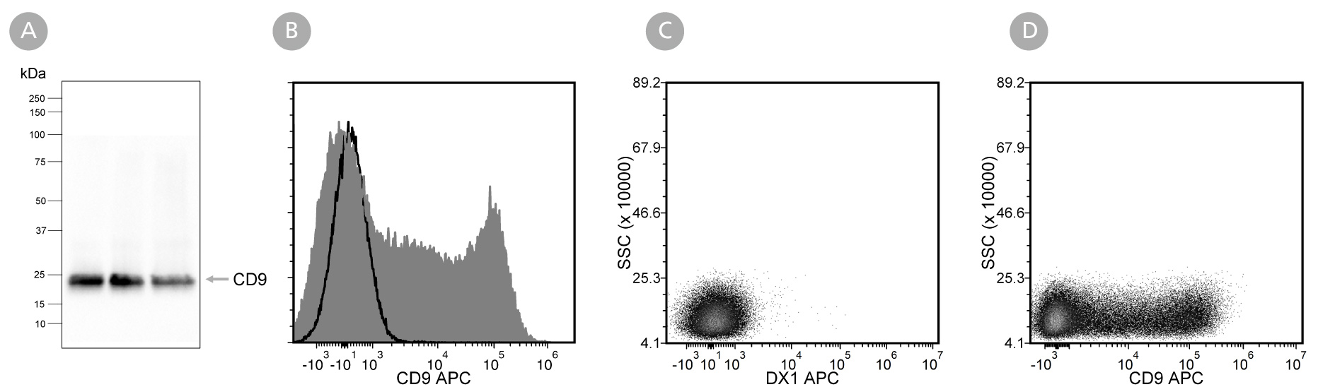
Figure 1. Analysis of Anti-Human CD9 Antibody, Clone HI9a with Western Blot and Flow Cytometry
Extracellular vesicles (EVs) were isolated from mesenchymal stromal cell (MSC)-conditioned medium using a 2 mL EV size exclusion chromatography column and analyzed by (A) western blot with Anti-Human CD9 Antibody, Clone HI9a. Lanes (left to right) were loaded with isolated fractions 9, 10, and 11, respectively. Further validation was performed by flow cytometry analysis of human peripheral blood mononuclear cells (PBMCs; viable lymphocytes gated for analysis) stimulated with phorbol myristate acetate (PMA) and ionomycin and then (B) labeled with Anti-Human CD9 Antibody, Clone HI9a, followed by a goat anti-mouse IgG1 antibody, APC (filled histogram), or Anti-Dextran Antibody, Clone DX1 (Catalog #60026) as an isotype control, followed by a goat anti-mouse IgG1 antibody, APC (solid line histogram), or (C) labeled with Anti-Dextran Antibody, Clone DX1 (Catalog #60026) as an isotype control, followed by a goat anti-mouse IgG1 antibody, APC, or (D) labeled with Anti-Human CD9 Antibody, Clone HI9a, followed by a goat anti-mouse IgG1 antibody, APC.