Anti-Human CD3 Antibody, Clone UCHT1
This antibody clone has been verified for purity assessments of cells isolated with EasySep™ kits, including EasySep™ Direct Human T Cell Isolation Kit (Catalog #19661), EasySep™ Human CD3 Positive Selection Kit II (Catalog #17851; partial blocking may be observed), EasySep™ HLA Whole Blood T Cell Enrichment Kit (Catalog #19951HLA) and EasySep™ HLA Whole Blood CD2 Positive Selection Kit (Catalog #18687HLA).
Data
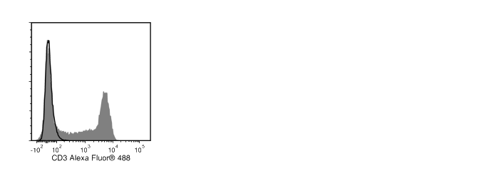
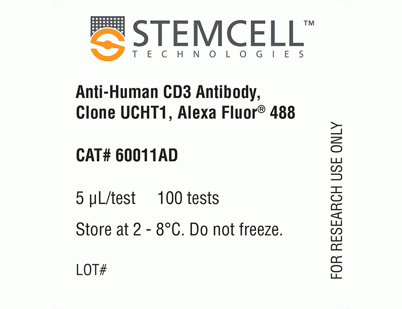
Figure 1. Data for Alexa Fluor® 488-Conjugated
Flow cytometry analysis of human peripheral blood mononuclear cells (PBMCs) labeled with Anti-Human CD3 Antibody, Clone UCHT1, Alexa Fluor® 488 (filled histogram) or a mouse IgG1, kappa Alexa Fluor® 488 isotype control antibody (solid line histogram).
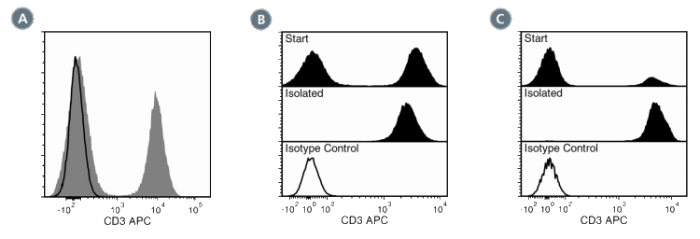
Figure 2. Data for APC-Conjugated
(A) Flow cytometry analysis of human peripheral blood mononuclear cells (PBMCs) labeled with Anti-Human CD3 Antibody, Clone UCHT1, APC (filled histogram) or a mouse IgG1, kappa APC isotype control antibody (black line histogram).
(B) Flow cytometry analysis of human buffy coat nucleated cells processed with the EasySep™ HLA Whole Blood CD3 Positive Selection Kit and labeled with Anti-Human CD3 Antibody, Clone UCHT1, APC. Histograms show labeling of buffy coat nucleated cells (Start) and isolated cells (Isolated). Labeling of start cells with a mouse IgG1, kappa APC isotype control antibody is shown (open histogram).
(C) Flow cytometry analysis of human whole blood nucleated cells processed with the EasySep™ HLA Whole Blood T Cell Enrichment Kit (Catalog #19951HLA). Histograms show labeling of HetaSep™-treated whole blood cells (Start) and isolated cells (Isolated). Labeling of start cells with a mouse IgG1, kappa APC isotype control antibody is shown (open histogram).
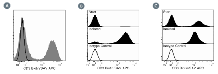
Figure 3. Data for Biotin-Conjugated
(A) Flow cytometry analysis of human peripheral blood mononuclear cells (PBMCs) labeled with Anti-Human CD3 Antibody, Clone UCHT1, Biotin followed by streptavidin (SAV) APC (filled histogram) or a mouse IgG1, kappa biotin isotype control antibody followed by SAV APC (black line histogram).
(B) Flow cytometry analysis of human buffy coat nucleated cells processed with the EasySep™ HLA Whole Blood CD2 Positive Selection Kit and labeled with Anti-Human CD3 Antibody, Clone UCHT1, Biotin followed by streptavidin (SAV) APC. Histograms show labeling of buffy coat nucleated cells (Start) and isolated cells (Isolated). Labeling of start cells with a mouse IgG1, kappa biotin isotype control antibodyfollowed by SAV APC is shown (open histogram).
(C) Flow cytometry analysis of human buffy coat nucleated cells processed with the EasySep™ HLA Whole Blood CD3 Positive Selection Kit and labeled with Anti-Human CD3 Antibody, Clone UCHT1, Biotin followed by streptavidin (SAV) APC. Histograms show labeling of buffy coat nucleated cells (Start) and isolated cells (Isolated). Labeling of start cells with a mouse IgG1, kappa biotin isotype control antibodyfollowed by SAV APC is shown (open histogram).
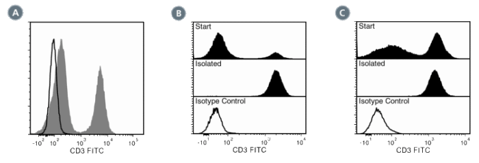
Figure 4. Data for FITC-Conjugated
(A) Flow cytometry analysis of human peripheral blood mononuclear cells (PBMCs) labeled with Anti-Human CD3 Antibody, Clone UCHT1, FITC (filled histogram) or a mouse IgG1, kappa FITC isotype control antibody (black line histogram).
(B) Flow cytometry analysis of human whole blood nucleated cells processed with the EasySep™ HLA Whole Blood T Cell Enrichment Kit and labeled with Anti-Human CD3 Antibody, Clone UCHT1, FITC. Histograms show labeling of HetaSep™-treated whole blood cells (Start) and isolated cells (Isolated). Labeling of start cells with a mouse IgG1, kappa FITC isotype control antibody is shown (open histogram).
(C) Flow cytometry analysis of human buffy coat nucleated cells processed with the EasySep™ HLA Whole Blood CD3 Positive Selection Kit and labeled with Anti-Human CD3 Antibody, Clone UCHT1, FITC. Histograms show labeling of buffy coat nucleated cells (Start) and isolated cells (Isolated). Labeling of start cells with a mouse IgG1, kappa FITC isotype control antibody is shown (open histogram).
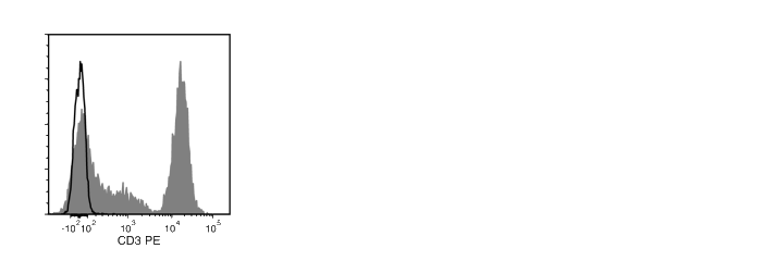
Figure 5. Data for PE-Conjugated
Flow cytometry analysis of human peripheral blood mononuclear cells (PBMCs) labeled with Anti-Human CD3 Antibody, Clone UCHT1, PE (filled histogram) or a mouse IgG1, kappa PE isotype control antibody (solid line histogram).
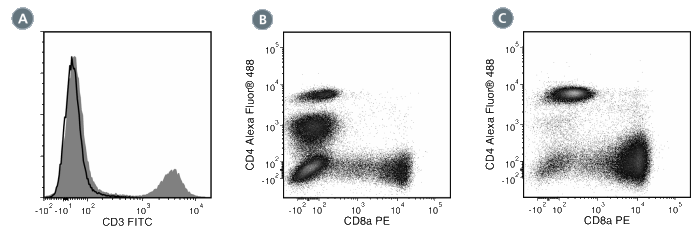
Figure 6. Data for Unconjugated
(A) Flow cytometry analysis of human peripheral blood mononuclear cells (PBMCs) labeled with Anti-Human CD3 Antibody, Clone UCHT1, followed by goat anti-mouse IgG, FITC (filled histogram). Labeling with a mouse IgG1, kappa isotype control antibody followed by goat anti-mouse IgG, FITC is shown (solid line histogram).
(B) Flow cytometry analysis of PBMCs prior to being processed with EasySep™ Human CD3 Positive Selection Kit (Catalog #18051), which uses Anti-Human CD3 Antibody, Clone UCHT1, to select CD3+ cells. The cells were labeled with Anti-Human CD4 Antibody, Clone OKT4, Alexa Fluor® 488 (Catalog #60016AD) and Anti-Human CD8 Antibody, Clone RPA-T8, PE (Catalog #60022PE).
(C) Flow cytometry analysis of PBMCs processed with EasySep™ Human CD3 Positive Selection Kit and labeled with Anti-Human CD4 Antibody, Clone OKT4, Alexa Fluor® 488 (Catalog #60016AD) and Anti-Human CD8 Antibody, Clone RPA-T8 (Catalog #60022PE).
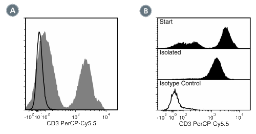
Figure 7. Data for PerCP-Cy55-Conjugated
(A) Flow cytometry analysis of human peripheral blood mononuclear cells (PBMCs) labeled with Anti-Human CD3 Antibody, Clone UCHT1, PerCP-Cy5.5 (filled histogram) or a mouse IgG1, kappa isotype control antibody, PerCP-Cy5.5 (solid line histogram).
(B) Flow cytometry analysis of human PBMCs processed with the EasySep™ Human CD3 Positive Selection Kit (Catalog #18051) and labeled with Anti-Human CD3 Antibody, Clone UCHT1, PerCP-Cy5.5. Histograms show labeling of PBMCs (Start) and isolated cells (Isolated). Labeling of start cells with a mouse IgG1, kappa isotype control antibody, PerCP-Cy5.5 is shown in the bottom panel (solid line histogram).
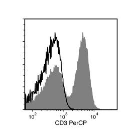
Figure 8. Data for PerCP-Conjugated
Flow cytometry analysis of human peripheral blood mononuclear cells (PBMCs) labeled with Anti-Human CD3 Antibody, Clone UCHT1, PerCP (filled histogram) or a mouse IgG1, kappa isotype control antibody, PerCP (solid line histogram).

