
Anti-Human CD3 Antibody, Clone OKT3


| Document Type | 产品名称 | Catalog # | Lot # | 语言 |
|---|---|---|---|---|
| Product Information Sheet | Anti-Human CD3 Antibody, Clone OKT3, FITC | 100-0285, 100-0286 | All | English |
| Product Information Sheet | Anti-Human CD3 Antibody, Clone OKT3 | 100-0287 | All | English |
| Product Information Sheet | Anti-Human CD3 Antibody, Clone OKT3, APC | 100-0288, 100-0289 | All | English |
| Safety Data Sheet | Anti-Human CD3 Antibody, Clone OKT3, FITC | 100-0285, 100-0286 | All | English |
| Safety Data Sheet | Anti-Human CD3 Antibody, Clone OKT3 | 100-0287 | All | English |
| Safety Data Sheet | Anti-Human CD3 Antibody, Clone OKT3, APC | 100-0288, 100-0289 | All | English |
Data
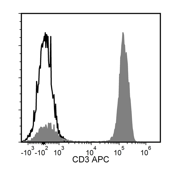
Figure 1. Data for Anti-Human CD3 Antibody, Clone OKT3, APC
Flow cytometry analysis of human peripheral blood mononuclear cells (PBMCs) labeled with Anti-Human CD3 Antibody, Clone OKT3, APC (filled histogram) or a mouse IgG2a, kappa APC isotype control antibody (solid line histogram). Viable lymphocytes were gated for analysis.
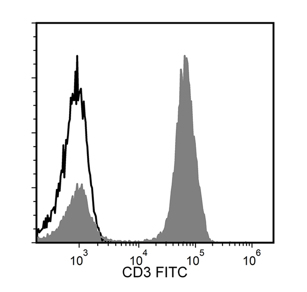
Figure 2. Data for Anti-Human CD3 Antibody, Clone OKT3, FITC
Flow cytometry analysis of human PBMCs labeled with Anti-Human CD3 Antibody, Clone OKT3, FITC (filled histogram) or a mouse IgG2a, kappa FITC isotype control antibody (solid line histogram). Viable lymphocytes were gated for analysis.
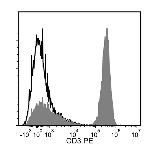
Figure 3. Data for Anti-Human CD3 Antibody, Clone OKT3, Unconjugated
Flow cytometry analysis of human PBMCs labeled with Anti-Human CD3 Antibody, Clone OKT3, followed by a goat anti-mouse IgG2a antibody, PE (filled histogram) or a mouse IgG2a, kappa isotype control antibody, followed by a goat anti-mouse IgG2a antibody, PE (solid line histogram). Viable lymphocytes were gated for analysis.