
Anti-Human CD2 Antibody, Clone RPA-2.10


Data
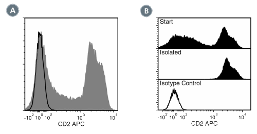
Figure 1. Data for Anti-Human CD2 Antibody, Clone RPA-2.10, APC
(A) Flow cytometry analysis of human buffy coat cells labeled with Anti-Human CD2 Antibody, Clone RPA-2.10, APC (filled histogram) or a mouse IgG1, kappa APC isotype control antibody (black line histogram).
(B) Flow cytometry analysis of human peripheral blood mononuclear cells (PBMCs) processed with the EasySep™ HLA CD3 Positive Selection Kit and labeled with Anti-Human CD2 Antibody, Clone RPA-2.10, APC. Histograms show labeling of PBMCs (Start) and isolated cells (Isolated). Labeling of start cells with a mouse IgG1, kappa APC isotype control antibody is shown (open histogram).
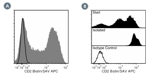
Figure 2. Data for Anti-Human CD2 Antibody, Clone RPA-2.10, Biotin
(A) Flow cytometry analysis of human buffy coat cells labeled with Anti-Human CD2 Antibody, Clone RPA-2.10, Biotin followed by streptavidin (SAV) APC (filled histogram) or a mouse IgG1, kappa biotin isotype control antibody followed by SAV APC (black line histogram).
(B) Flow cytometry analysis of human peripheral blood mononuclear cells (PBMCs) processed with the EasySep™ HLA CD3 Positive Selection Kit and labeled with Anti-Human CD2 Antibody, Clone RPA-2.10, Biotin followed by streptavidin (SAV) APC. Histograms show labeling of PBMCs (Start) and isolated cells (Isolated). Labeling of start cells with a mouse IgG1, kappa biotin isotype control antibody followed by SAV APC is shown (open histogram).
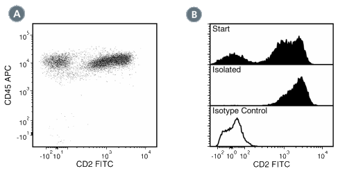
Figure 3. Data for Anti-Human CD2 Antibody, Clone RPA-2.10, FITC
(A) Flow cytometry analysis of human peripheral blood mononuclear cells (PBMCs) labeled with Anti-Human CD2 Antibody, Clone RPA-2.10, FITC and anti-human CD45 APC.
(B) Flow cytometry analysis of human PBMCs processed with the EasySep™ HLA CD3 Positive Selection Kit and labeled with Anti-Human CD2 Antibody, Clone RPA-2.10, FITC. Histograms show labeling of PBMCs (Start) and isolated cells (Isolated). Labeling of start cells with a mouse IgG1, kappa FITC isotype control antibody is shown (open histogram).
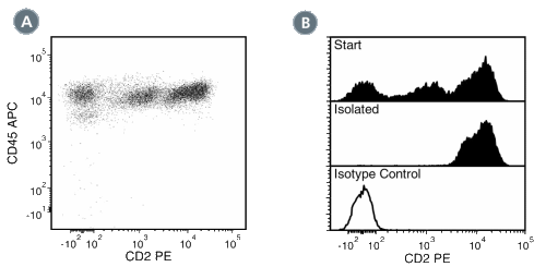
Figure 4. Data for Anti-Human CD2 Antibody, Clone RPA-2.10, PE
(A) Flow cytometry analysis of human peripheral blood mononuclear cells (PBMCs) labeled with Anti-Human CD2 Antibody, Clone RPA-2.10, PE and anti-human CD45 APC. (B) Flow cytometry analysis of human PBMCs processed with the EasySep™ HLA CD3 Positive Selection Kit and labeled with Anti-Human CD2 Antibody, Clone RPA-2.10, PE. Histograms show labeling of PBMCs (Start) and isolated cells (Isolated). Labeling of start cells with a mouse IgG1, kappa PE isotype control antibody is shown (open histogram).
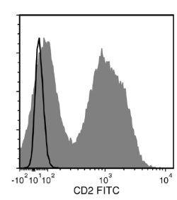
Figure 5. Data for Anti-Human CD2 Antibody, Clone RPA-2.10
Flow cytometry analysis of human peripheral blood mononuclear cells (PBMCs) labeled with Anti-Human CD2 Antibody, Clone RPA-2.10, followed by Goat Anti-Mouse IgG (H+L) Antibody, Polyclonal, FITC (Catalog #60138FI; filled histogram), or a mouse IgG1, kappa isotype control antibody followed by Goat Anti-Mouse IgG (H+L) Antibody, Polyclonal, FITC (solid line histogram).
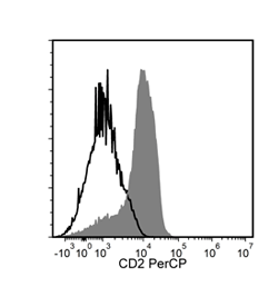
Figure 6. Data for Anti-Human CD2 Antibody, Clone RPA-2.10, PerCP
Flow cytometry analysis of human peripheral blood mononuclear cells (PBMCs) labeled with Anti-Human CD2 Antibody, Clone RPA-2.10, PerCP (filled histogram) or a mouse IgG1, kappa isotype control antibody, PerCP (solid line histogram).