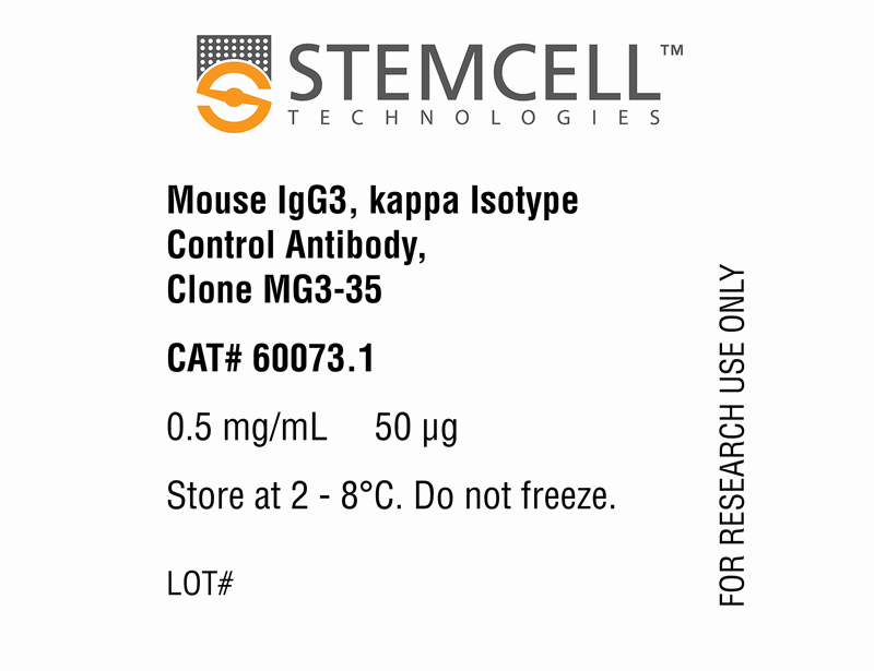
Mouse IgG3, kappa Isotype Control Antibody, Clone MG3-35
This antibody clone has been verified for use as an isotype control antibody for assessing non-specific binding to cells in applications such as flow cytometry and immunofluorescence microscopy applications (surface and intracellular staining).
Data
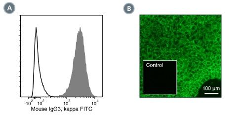
Figure 1. Data for Unconjugated
(A) Flow cytometry analysis of human iPS cells labeled with Mouse IgG3, kappa Isotype Control Antibody, Clone MG3-35, followed by a goat anti-mouse IgG antibody, FITC (solid line histogram). Filled histogram shows labeling with a mouse IgG3, kappa positive control antibody (Anti-Human SSEA-4 Antibody, Clone MC-813-70, Catalog #60062) followed by a goat anti-mouse IgG antibody, FITC. (B) Human ES cells were cultured in mTeSR™1 on Corning® Matrigel®-coated glass slides, then fixed and stained with Anti-Human SSEA-4 Antibody, Clone MC-813-70 (Catalog #60062), followed by goat anti-mouse IgG, FITC. Inset shows cells labeled with Mouse IgG3, kappa Isotype Control Antibody, Clone MG3-35.
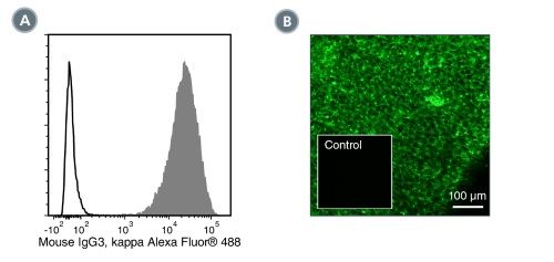
Figure 2. Data for Alexa Fluor® 488-Conjugated
(A) Flow cytometry analysis of human iPS cells labeled with Mouse IgG3, kappa Isotype Control Antibody, Clone MG3-35, Alexa Fluor® 488 (solid line histogram). Filled histogram shows labeling with a mouse IgG3, kappa positive control antibody (Anti-Human SSEA-4 Antibody, Clone MC-813-70, Alexa Fluor® 488, Catalog #60062AD). B) Human ES cells were cultured in mTeSR™1 on Corning Matrigel®-coated glass slides, then fixed and stained with Anti-Human SSEA-4 Antibody, Clone MC-813-70, Alexa Fluor® 488. Inset shows cells labeled with Mouse IgG3, kappa Isotype Control Antibody, Clone MG3-35, Alexa Fluor® 488.
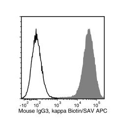
Figure 3. Data for Biotin-Conjugated
Flow cytometry analysis of human ES cells labeled with Mouse IgG3, kappa Isotype Control Antibody, Clone MG3-35, Biotin followed by streptavidin (SAV) APC (solid line histogram). Filled histogram shows labeling with a mouse IgG3, kappa positive control antibody (Anti-Human SSEA-4 Antibody, Clone MC-813-70, Biotin, Catalog #60062BT) followed by SAV APC.
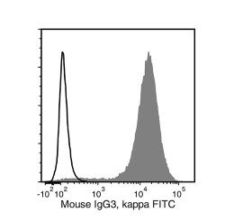
Figure 4. Data for FITC-Conjugated
Flow cytometry analysis of human ES cells labeled with Mouse IgG3, kappa Isotype Control Antibody, Clone MG3-35, FITC (solid line histogram). Filled histogram shows labeling with a mouse IgG3, kappa positive control antibody (Anti-Human SSEA-4 Antibody, Clone MC-813-70, FITC, Catalog #60062FI).
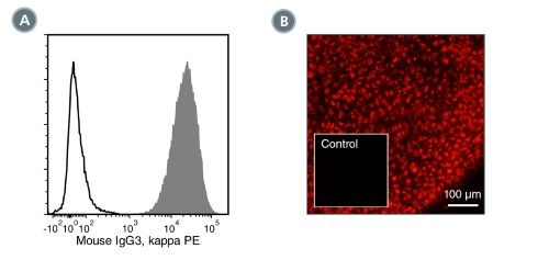
Figure 5. Data for PE-Conjugated
(A) Flow cytometry analysis of human iPS cells labeled with Mouse IgG3, kappa Isotype Control Antibody, Clone MG3-35, PE (solid line histogram). Filled histogram shows labeling with a mouse IgG3, kappa positive control antibody (Anti-Human SSEA-4 Antibody, Clone MC-813-70, PE, Catalog #60062PE). (B) Human ES cells were cultured in mTeSR™1 on Corning® Matrigel®-coated glass slides, then fixed and stained with Anti-Human SSEA-4 Antibody, Clone MC-813-70, PE (Catalog #60062PE). Inset shows cells labeled with Mouse IgG3, kappa Isotype Control Antibody, Clone MG3-35.

