STEMdiff™ Hematopoietic Kit

For differentiation of human ES or iPS cells into hematopoietic progenitor cells
概要
STEMdiff™ Hematopoietic Kit includes a serum-free basal medium and supplements for the feeder-free differentiation of human embryonic stem (ES) and induced pluripotent stem (iPS) cells into hematopoietic progenitor cells expressing CD34, CD45, and CD43.

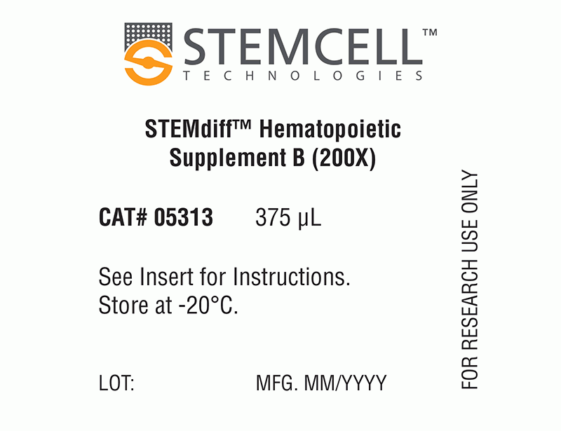
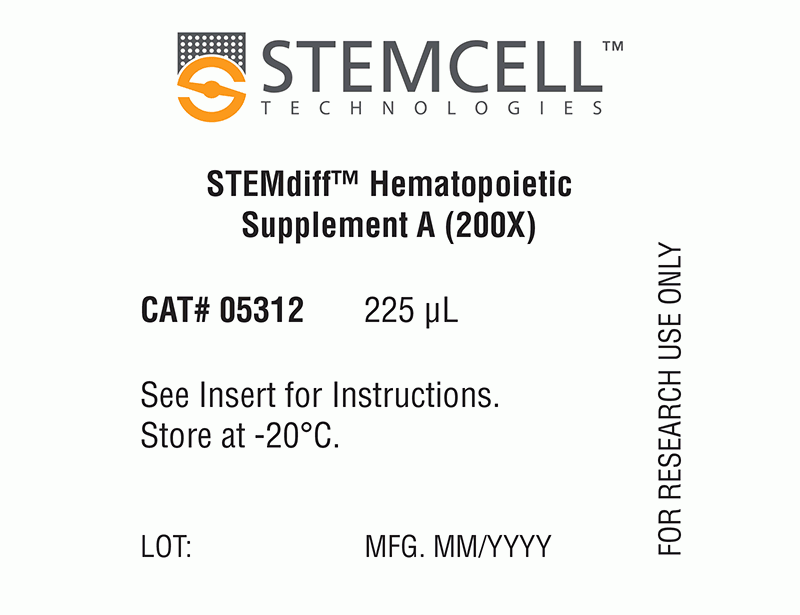
The simple, 12-day differentiation protocol is performed in two stages. During the first 3 days, STEMdiff™ Hematopoietic Supplement A is added to the basal medium to induce cells toward mesoderm. For the subsequent 9 days, mesodermal cells are further differentiated into hematopoietic progenitor cells using basal medium supplemented with STEMdiff™ Hematopoietic Supplement B. At the end of the 12-day protocol, hematopoietic cells can be easily harvested from the culture supernatant. This population typically contains 25 - 65% (average 43%) CD34+CD45+ progenitor cells, including progenitor cells that have the capacity to form hematopoietic colonies in the colony-forming unit (CFU) assay.

STEMdiff™ Hematopoietic Kit has been optimized for differentiation of cells maintained in mTeSR™1 (Catalog #85850), mTeSR™ Plus(Catalog #100-0276), or TeSR™-E8™ (Catalog #05990).
Components
• STEMdiff™ Hematopoietic Basal Medium, 120 mL
• STEMdiff™ Hematopoietic Supplement A (200X), 225 µL
• STEMdiff™ Hematopoietic Supplement B (200X), 375 µL
 
Subtype
Specialized Media
Cell Type
Hematopoietic Stem and Progenitor Cells, Pluripotent Stem Cells
Species
Human
Application
Cell Culture, Differentiation
Brand
STEMdiff
Area of Interest
Stem Cell Biology
Formulation
Serum-Free
数据及文献

Data

Hematopoietic Cell Differentiation Protocol

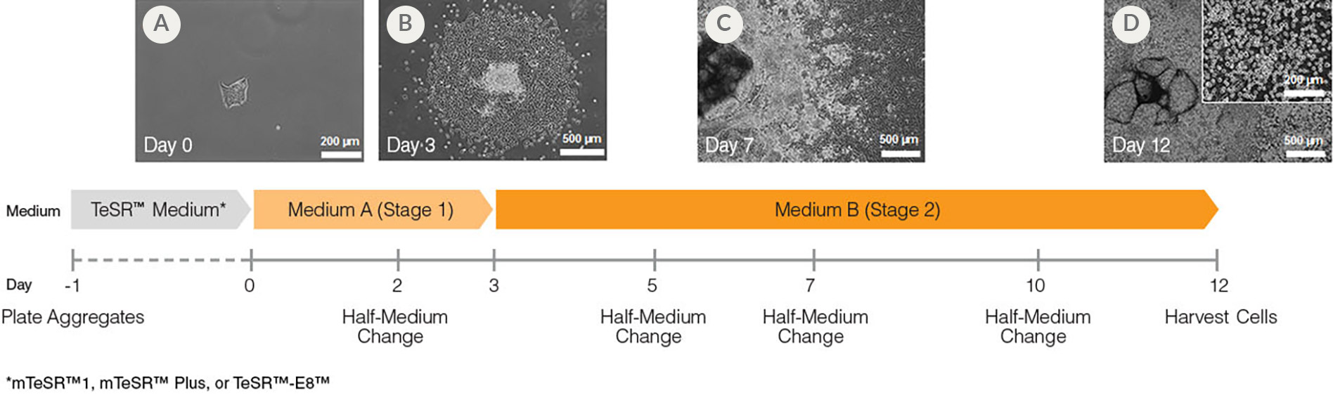
Figure 1. Hematopoietic Differentiation Protocol

On Day -1, harvest and seed human ES/iPS cell colonies as small aggregates in mTeSR™1, mTeSR™ Plus, or TeSR™-E8. After one day, TeSR™ medium is replaced with Medium A (STEMdiff™ Hematopoietic Basal Medium containing Supplement A) to begin inducing the cells towards a mesoderm-like state (day 0). On day 2, a half medium change is performed with fresh Medium A. On day 3, the medium is changed to Medium B (STEMdiff™ Hematopoietic Basal Medium containing Supplement B) with half medium changes on days 5, 7 and 10, to promote further differentiation into hematopoietic cells. Typically, by day 12, large numbers of HPCs can be harvested from the culture supernatant.

Morphology of hPSC-Derived HPCs

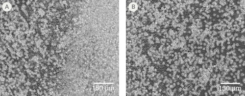
Figure 2. Morphology of hPSC-Derived HPCs

Representative images of (A) hES (H1) cells and (B) hiPS (WLS-1C) cells on Day 12 of differentiation to HPCs using the STEMdiff™ Hematopoietic Kit. Differentiated cells exhibit typical HPC morphology as round cells that float freely in suspension.

Efficient and Robust Generation of CD34+CD45+/CD43+ HPCs

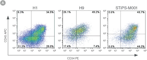
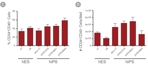
Figure 3. Efficient and Robust Generation of CD34+CD45+/CD43+ HPCs

hES and hiPS cells were cultured for 12 days in single wells of 12-well plates using the STEMdiff™ Hematopoietic Kit. At the end of the culture period, cells in suspension were harvested and analyzed by flow cytometry for expression of hematopoietic cell surface markers: CD34, CD45 and CD43. (A,B) Example flow cytometry plots for hematopoietic cell surface-marker analysis of cultures of hES (H1 and H9) and hiPS (STiPS-M001) cells. (C,D) Percentages and total numbers of CD34+CD45+ cells in cultures of hES (H1 and H9) or hiPS (WLS-1C, STiPS-F016, STiPS-M001 and STiPS-B004) cells are shown. Data shown as mean ± SEM; n ≥ 3.

hPSC-Derived HPCs Produce Colonies of Multiple Lineages

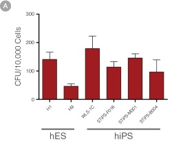
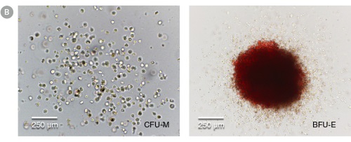
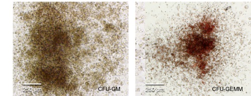
Figure 4. hPSC-Derived HPCs Produce Colonies of Multiple Lineages

Cells in suspension were harvested from the cultures on Day 12 of the hematopoietic differentiation protocol and assessed in colony-forming unit (CFU) assays using MethoCult™ H4435 Enriched (Catalog #04435) methylcellulose-based medium. (A) CFU frequencies in cultures of 6 different hPSC lines. (Data shown as mean ± SEM; n ≥ 3.) The CFU frequencies were variable between the different cell lines, with on average approximately 120 CFU per 10,000 hPSC-derived HPCs plated. (B) The progenitor cell types observed included granulocyte/macrophage (CFU-M, CFU-G and CFU-GM), erythroid (BFU-E and CFU-E) and occasional mixed (CFU-GEMM) colonies. Representative colony images are shown at 40X magnification.

Density plots showing CD34+ and CD45+ expression and percentage of cells co-expressing CD34+ and CD45+ and graphs showing total number of viable cells harvested.

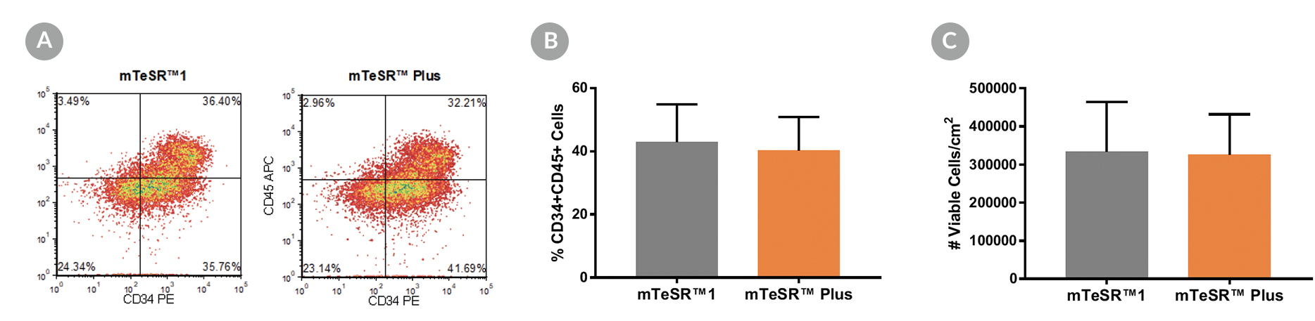
Figure 5. Generation of Hematopoietic Progenitor Cells from hPSCs Maintained in mTeSR™ Plus

Human ES (H1, H9) and iPS (STiPS-M001, WLS-1C) cell lines maintained in mTeSR™1 (daily feeds) or mTeSR™ Plus (restricted feeds) were differentiated to hematopoietic progenitor cells using the STEMdiff™ Hematopoietic Kit. At the end of the differentiation period, cells were harvested from the supernatant and analyzed by flow cytometry for co-expression of CD34+ and CD45+ . (A) Representative density plots showing CD34+ and CD45+ expression, (B) percentage of cells co-expressing CD34+ and CD45+ , and (C) total number of viable cells harvested are shown. Data are expressed as the mean (± SEM); n=4.

Publications (3)

Journal of virology 2021 jan CD34+ Hematopoietic Progenitor Cell Subsets Exhibit Differential Ability To Maintain Human Cytomegalovirus Latency and Persistence. L. B. Crawford et al.

Abstract

In human cytomegalovirus (HCMV)-seropositive patients, CD34+ hematopoietic progenitor cells (HPCs) provide an important source of latent virus that reactivates following cellular differentiation into tissue macrophages. Multiple groups have used primary CD34+ HPCs to investigate mechanisms of viral latency. However, analyses of mechanisms of HCMV latency have been hampered by the genetic variability of CD34+ HPCs from different donors, availability of cells, and low frequency of reactivation. In addition, multiple progenitor cell types express surface CD34, and the frequencies of these populations differ depending on the tissue source of the cells and culture conditions in vitro In this study, we generated CD34+ progenitor cells from two different embryonic stem cell (ESC) lines, WA01 and WA09, to circumvent limitations associated with primary CD34+ HPCs. HCMV infection of CD34+ HPCs derived from either WA01 or WA09 ESCs supported HCMV latency and induced myelosuppression similar to infection of primary CD34+ HPCs. Analysis of HCMV-infected primary or ESC-derived CD34+ HPC subpopulations indicated that HCMV was able to establish latency and reactivate in CD38+ CD90+ and CD38+/low CD90- HPCs but persistently infected CD38- CD90+ cells to produce infectious virus. These results indicate that ESC-derived CD34+ HPCs can be used as a model for HCMV latency and that the virus either latently or persistently infects specific subpopulations of CD34+ cells.IMPORTANCE Human cytomegalovirus infection is associated with severe disease in transplant patients and understanding how latency and reactivation occur in stem cell populations is essential to understand disease. CD34+ hematopoietic progenitor cells (HPCs) are a critical viral reservoir; however, these cells are a heterogeneous pool with donor-to-donor variation in functional, genetic, and phenotypic characteristics. We generated a novel system using embryonic stem cell lines to model HCMV latency and reactivation in HPCs with a consistent cellular background. Our study defined three key stem cell subsets with differentially regulated latent and replicative states, which provide cellular candidates for isolation and treatment of transplant-mediated disease. This work provides a direction toward developing strategies to control the switch between latency and reactivation.
Stem cell reports 2020 feb iPSC-Based Modeling of RAG2 Severe Combined Immunodeficiency Reveals Multiple T Cell Developmental Arrests. M. Themeli et al.

Abstract

RAG2 severe combined immune deficiency (RAG2-SCID) is a lethal disorder caused by the absence of functional T and B cells due to a differentiation block. Here, we generated induced pluripotent stem cells (iPSCs) from a RAG2-SCID patient to study the nature of the T cell developmental blockade. We observed a strongly reduced capacity to differentiate at every investigated stage of T cell development, from early CD7-CD5- to CD4+CD8+. The impaired differentiation was accompanied by an increase in CD7-CD56+CD33+ natural killer (NK) cell-like cells. T cell receptor D rearrangements were completely absent in RAG2SCID cells, whereas the rare T cell receptor B rearrangements were likely the result of illegitimate rearrangements. Repair of RAG2 restored the capacity to induce T cell receptor rearrangements, normalized T cell development, and corrected the NK cell-like phenotype. In conclusion, we succeeded in generating an iPSC-based RAG2-SCID model, which enabled the identification of previously unrecognized disorder-related T cell developmental roadblocks.
Molecular neurodegeneration 2018 DEC Development and validation of a simplified method to generate human microglia from pluripotent stem cells. A. McQuade et al.

Abstract

BACKGROUND Microglia, the principle immune cells of the brain, play important roles in neuronal development, homeostatic function and neurodegenerative disease. Recent genetic studies have further highlighted the importance of microglia in neurodegeneration with the identification of disease risk polymorphisms in many microglial genes. To better understand the role of these genes in microglial biology and disease, we, and others, have developed methods to differentiate microglia from human induced pluripotent stem cells (iPSCs). While the development of these methods has begun to enable important new studies of microglial biology, labs with little prior stem cell experience have sometimes found it challenging to adopt these complex protocols. Therefore, we have now developed a greatly simplified approach to generate large numbers of highly pure human microglia. RESULTS iPSCs are first differentiated toward a mesodermal, hematopoietic lineage using commercially available media. Highly pure populations of non-adherent CD43+ hematopoietic progenitors are then simply transferred to media that includes three key cytokines (M-CSF, IL-34, and TGF$\beta$-1) that promote differentiation of homeostatic microglia. This updated approach avoids the prior requirement for hypoxic incubation, complex media formulation, FACS sorting, or co-culture, thereby significantly simplifying human microglial generation. To confirm that the resulting cells are equivalent to previously developed iPSC-microglia, we performed RNA-sequencing, functional testing, and transplantation studies. Our findings reveal that microglia generated via this simplified method are virtually identical to iPS-microglia produced via our previously published approach. To also determine whether a small molecule activator of TGF$\beta$ signaling (IDE1) can be used to replace recombinant TGF$\beta$1, further reducing costs, we examined growth kinetics and the transcriptome of cells differentiated with IDE1. These data demonstrate that a microglial cell can indeed be produced using this alternative approach, although transcriptional differences do occur that should be considered. CONCLUSION We anticipate that this new and greatly simplified protocol will enable many interested labs, including those with little prior stem cell or flow cytometry experience, to generate and study human iPS-microglia. By combining this method with other advances such as CRISPR-gene editing and xenotransplantation, the field will continue to improve our understanding of microglial biology and their important roles in human development, homeostasis, and disease.
View All Publications
Top